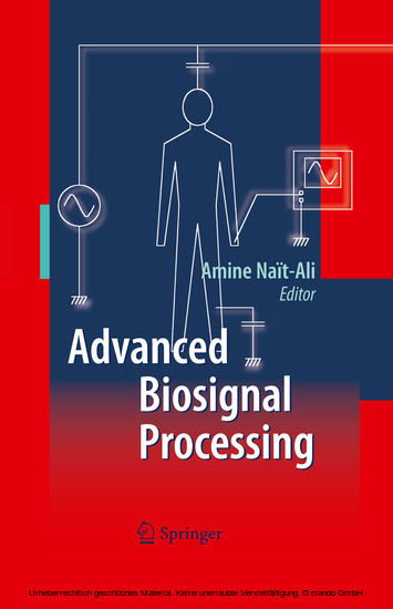
Advanced Biosignal Processing
Generally speaking, Biosignals refer to signals recorded from the human body. They can be either electrical (e. g. Electrocardiogram (ECG), Electroencephalogram (EEG), Electromyogram (EMG), etc. ) or non-electrical (e. g. breathing, movements, etc. ). The acquisition and processing of such signals play an important role in clinical routines. They are usually considered as major indicators which provide clinicians and physicians with useful information during diagnostic and monitoring processes. In some applications, the purpose is not necessarily medical. It may also be industrial. For instance, a real-time EEG system analysis can be used to control and analyze the vigilance of a car driver. In this case, the purpose of such a system basically consists of preventing crash risks. Furthermore, in certain other appli- tions,asetof biosignals (e. g. ECG,respiratorysignal,EEG,etc. ) can be used toc- trol or analyze human emotions. This is the case of the famous polygraph system, also known as the 'lie detector', the ef ciency of which remains open to debate! Thus when one is dealing with biosignals, special attention must be given to their acquisition, their analysis and their processing capabilities which constitute the nal stage preceding the clinical diagnosis. Naturally, the diagnosis is based on the information provided by the processing system.
1;Preface;5 2;Contents;10 3;Contributors;12 4;to 1 Biosignals: Acquisition and General Properties;16 4.1;1.1 Biopotential Recording;16 4.1.1;1.1.1 Biopotentials Recording Electrodes;17 4.1.1.1;1.1.1.1 Equivalent Circuit;17 4.1.1.2;1.1.1.2 Other Electrodes;17 4.1.2;1.1.2 Power Supply Artifact Rejection;18 4.1.2.1;1.1.2.1 Displacement Currents;18 4.1.2.2;1.1.2.2 Common Mode Rejection;20 4.1.2.3;1.1.2.3 Magnetic Induction;20 4.1.2.4;1.1.2.4 The Right Leg Driver;21 4.1.3;1.1.3 Safety;22 4.1.3.1;1.1.3.1 Current Limitation;22 4.1.3.2;1.1.3.2 Galvanic Isolation;22 4.1.4;1.1.4 To Conclude this Section...;22 4.2;1.2 General Properties of Some Common Biosignals;23 4.2.1;1.2.1 The Electrocardiogram (ECG);23 4.2.2;1.2.2 The Electroencephalogram (EEG);24 4.2.3;1.2.3 Evoked Potentials (EP);25 4.2.4;1.2.4 The Electromyogram (EMG);26 4.3;1.3 Some Comments;27 4.4;1.4 Conclusion;27 5;to 2 Extraction of ECG Characteristics Using Source Separation Techniques: Exploiting Statistical Independence and Beyond;29 5.1;2.1 Introduction;29 5.2;2.2 Approaches to Signal Extraction in the ECG;34 5.3;2.3 Blind Source Separation;35 5.3.1;2.3.1 Signal Model and Assumptions;35 5.3.2;2.3.2 Why Blind?;39 5.3.3;2.3.3 Achieving the Separation;40 5.4;2.4 Second-Order Approaches: Principal Component Analysis;41 5.4.1;2.4.1 Principle;41 5.4.2;2.4.2 Covariance Matrix Diagonalization;41 5.4.3;2.4.3 Limitations of Second-Order Statistics;42 5.5;2.5 Second-Order Approaches Exploiting Spectral Diversity;43 5.6;2.6 Higher-Order Approaches: Independent Component Analysis;45 5.6.1;2.6.1 Contrast Functions, Independence and Non-Gaussianity;45 5.6.2;2.6.2 Source Separation or Source Extraction?;47 5.6.3;2.6.3 Optimal Step-Size Iterative Search;49 5.7;2.7 Exploiting Prior Information;49 5.7.1;2.7.1 Source Statistical Characterization;50 5.7.1.1;2.7.1.1 Combining Non-Gaussianity and Spectral Features;51 5.7.1.2;2.7.1.2 Extraction of Sources with Known Kurtosis Sign;51 5.7.2;2.7.2 Reference Signal;53 5.7.3;2.7.3 Spatial Reference;55 5.8;2.8 Conclusions and Outlook;56 6;to 3 ECG Processing for Exercise Test;62 6.1;3.1 Introduction;62 6.2;3.2 ECG Acquisition During Exercise Test;64 6.3;3.3 Interval Estimation and Analysis;64 6.3.1;3.3.1 Heart Rate Variability Analysis;65 6.3.2;3.3.2 PR Interval Estimation;71 6.4;3.4 Shape Variations Estimation and Analysis;73 6.4.1;3.4.1 Curve Registration of P-waves During Exercise;74 6.4.1.1;3.4.1.1 Self-Modelling Registration (SMR);75 6.4.1.2;3.4.1.2 Application to Real P-waves;76 6.4.2;3.4.2 Simulation Study;76 6.5;3.5 Signal Averaging with Exercise;78 6.5.1;3.5.1 Equal Shape Signals;78 6.5.2;3.5.2 Non Equal Shape Signals;80 7;to 4 Statistical Models Based ECG Classification;83 7.1;4.1 Introduction;83 7.2;4.2 Hidden Markov Models;85 7.2.1;4.2.1 Overview;85 7.2.2;4.2.2 Heart Beat Modeling;87 7.2.3;4.2.3 Beat Classification;88 7.2.4;4.2.4 HMM for Beat Segmentation and Classification;90 7.2.4.1;4.2.4.1 Parameter Extraction;90 7.2.4.2;4.2.4.2 Training HMMs;91 7.2.4.3;4.2.4.3 Classifying Patterns;92 7.2.5;4.2.5 HMM Adaptation Adaptation ;93 7.2.6;4.2.6 Discussion;94 7.3;4.3 Hidden Markov Trees;95 7.3.1;4.3.1 Overview;95 7.3.2;4.3.2 Electrocardiogram Delineation by HMT;96 7.3.2.1;4.3.2.1 Model Training training ;97 7.3.2.2;4.3.2.2 Scale Segmentation segmentation ;98 7.3.2.3;4.3.2.3 Inter-Scale Fusion;99 7.3.2.4;4.3.2.4 Discussion;101 7.3.3;4.3.3 Association of HMM and HMT;102 7.4;4.4 Conclusions;103 8;to 5 Heart Rate Variability Time-Frequency Analysis for Newborn Seizure Detection;106 8.1;5.1 Identification of Newborn Seizures Using EEG, ECG and HRV Signals;106 8.1.1;5.1.1 Origin of Seizures;107 8.1.2;5.1.2 Seizure Manifestation;107 8.1.3;5.1.3 The Need for Early Seizure Detection;107 8.1.4;5.1.4 Seizure Monitoring Through EEG;108 8.1.5;5.1.5 Limitations of EEG-Based Seizure Identification;108 8.1.6;5.1.6 ECG-Based Seizure Identification;108 8.1.7;5.1.7 Combining EEG and ECG for R
Nait-Ali, Amine
| ISBN | 9783540895060 |
|---|---|
| Artikelnummer | 9783540895060 |
| Medientyp | E-Book - PDF |
| Copyrightjahr | 2009 |
| Verlag | Springer-Verlag |
| Umfang | 378 Seiten |
| Sprache | Englisch |
| Kopierschutz | Digitales Wasserzeichen |

