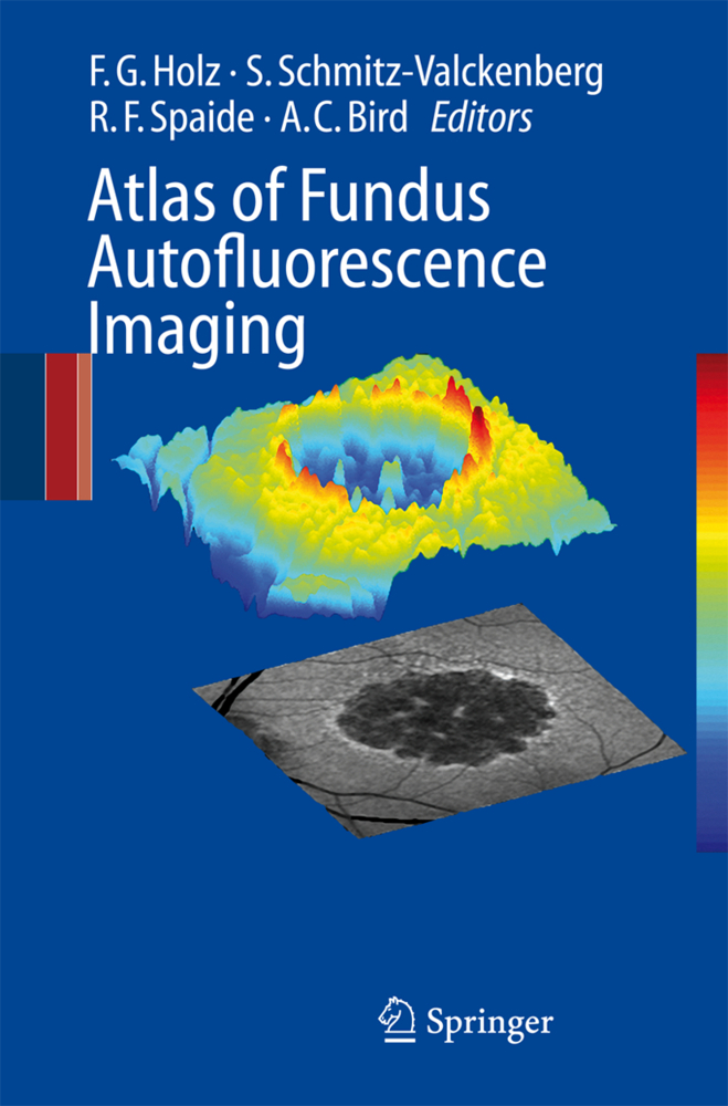
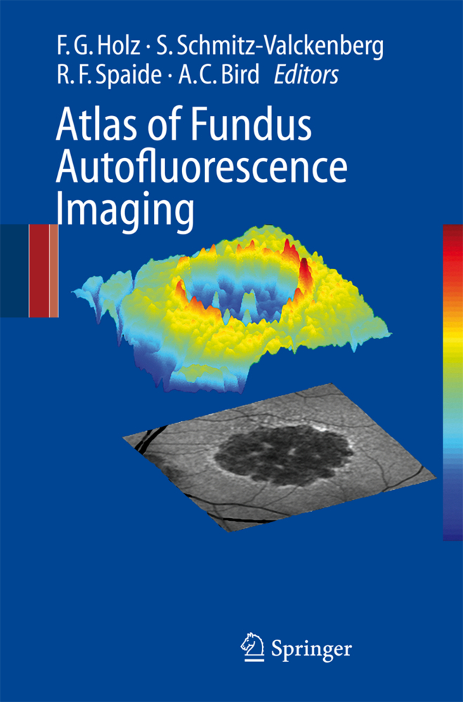
During recent years, FAF (Fundus autofluorescence) imaging has been shown to be useful in various retinal diseases with regard to diagnostics, documentation of changes, identification of disease progression, and monitoring of novel therapies. Hereby, FAF imaging gives additional information above and beyond conventional imaging tools.
This unique atlas provides a comprehensive and up-to-date overview of FAF imaging in retinal diseases. It also compares FAF findings with other imaging techniques such as
fundus photograph, fluorescein- and ICG angiography as well as optical coherence tomography.
General ophthalmologists as well as retina specialists will find this a very useful guide which illustrates typical FAF characteristics of various retinal diseases.
Methodology
Lipofuscin of the Retinal Pigment EpitheliumOrigin of Fundus Autofluorescence
Fundus Autofluorescence Imaging with the Confocal Scanning Laser Ophthalmoscope
How To Obtain the Optimal Fundus Autofluorescence Image with the Confocal Scanning Laser Ophthalmoscope
Autofluorescence Imaging with the Fundus Camera
Macular Pigment Measurement-Theoretical Background
Macular Pigment Measurement -Clinical Applications
Evaluation of Fundus Autofluorescence Images
Clinical Application
Macular and Retinal Dystrophies
Discrete Lines of Increased Fundus Autofluorescence in Various Forms of Retinal Dystrophies
Age-Related Macular Degeneration I-Early Manifestation
Age-Related Macular Degeneration II-Geographic Atrophy
Age-Related Macular Degeneration III-Pigment Epithelium Detachment
Age-Related Macular Degeneration IV-Choroidal Neovascularization (CNV)
Idiopathic Macular Telangiectasia
Chorioretinal Inflammatory Disorders
Autofluorescence from the Outer Retina and Subretinal Space
Miscellaneous
Perspectives in Imaging Technologies
Perspectives in Imaging Technologies.
Holz, Frank G
Schmitz-Valckenberg, Steffen
Spaide, Richard F.
Bird, Alan C.
| ISBN | 978-3-540-71993-9 |
|---|---|
| Artikelnummer | 9783540719939 |
| Medientyp | Buch |
| Copyrightjahr | 2007 |
| Verlag | Springer, Berlin |
| Umfang | XIII, 341 Seiten |
| Abbildungen | XIII, 341 p. |
| Sprache | Englisch |


