CT Teaching Manual
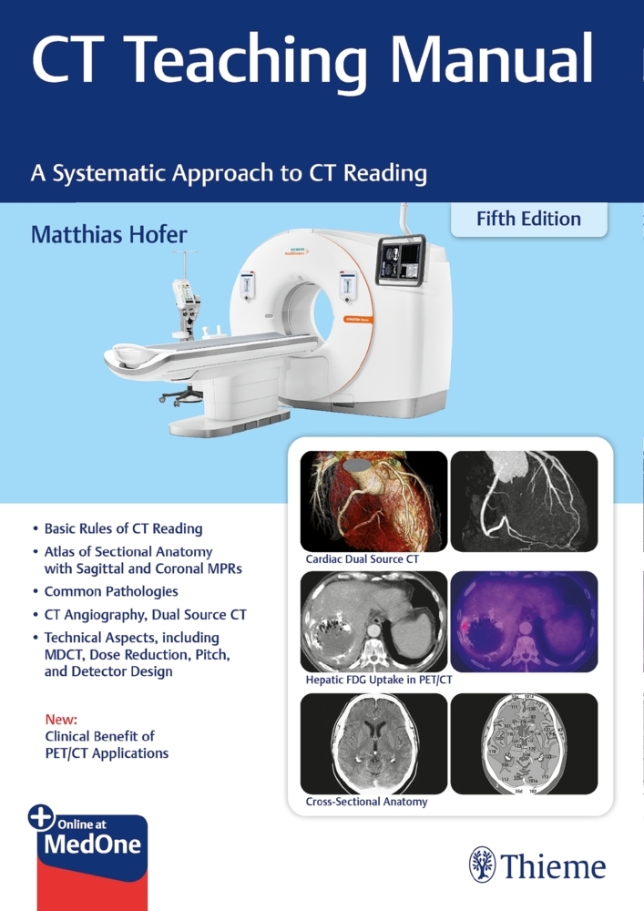
Ideal for residents starting in radiology and radiologic technologists, this concise manual is the perfect introduction to the physics and practice of CT and the interpretation of basic CT images. Designed as a systematic learning tool, it introduces the use of CT scanners for all organs, and includes positioning, use of contrast media, representative CT scans of normal and pathological findings, explanatory drawings with keyed anatomic structures, and an overview of the most important measurement data. Finally, self-assessment quizzes including answers at the end of each chapter help the reader monitor progress and evaluate knowledge gained.
New in this fifth edition: Recent technical developments such as dual source CT, protocols for CT angiography, and PET/CT fusion.
Physical and Technical Fundamentals
Basic Rules for Reading CT Examinations
Preparing the Patient
Administration of Contrast Agents
Cranial CT
Cranial CT: Normal Findings
Cranial Pathology
Cervical CT
Cervical Pathology
Chest CT
Chest CT Pathology
-Chest Wall
-Mediastinum
-Lung
Abdominal CT
Abdominal Pathology
-Abdominal Wall
-Liver
-Biliary Tract
-Gallbladder
-Spleen
-Pancreas
-Adrenal Glands
-Kidneys
-Urinary Bladder
-Reproductive Organs
-Gastrointestinal Tract
-Retroperitoneum
-Skeletal Changes
Spinal Column: Skeletal Pathology
Lower Extremity
Radiation Safety
CT Angiography
Contrast Injectors
Dual Source CT
Introduction to PET/CT
Anatomy in Coronal MPRs
Anatomy in Sagittal MPRs
Hofer, Matthias
| ISBN | 9783132442634 |
|---|---|
| Artikelnummer | 9783132442634 |
| Medientyp | Non Books |
| Auflage | 5. Aufl. |
| Copyrightjahr | 2021 |
| Verlag | Thieme, Stuttgart |
| Umfang | 232 Seiten |
| Abbildungen | 1243 Abb. |
| Sprache | Englisch |

