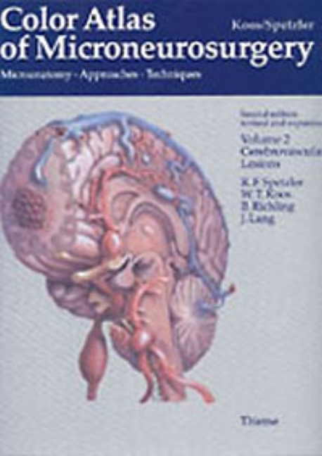
Color Atlas of Microneurosurgery: Volume 2 - Cerebrovascular Lesions
Color Atlas of Microneurosurgery: Volume 2 - Cerebrovascular Lesions
Refinements in the neurosurgical armamentarium continue to push the borders of neurosurgery forward. Lesions considered inoperable a few years ago can now be resected, especially in the region of the skull base. These new developments, plus rapid technological innovations in microneurosurgery, have dramatically altered the scope of modern neurosurgery.
Now, with Volume 2 of the acclaimed Color Atlas of Microneurosurgery, the distinguished authors provide detailed descriptions of surgical anatomy and the major neurosurgical approaches to cerebrovascular lesions. You will find coverage of aneurysms, arteriovenuous malformations, cerebrovascular malformations, and vascular compression all derived from a wide range of etiologies. Divided into three sections on anatomy, surgical approaches, and underlying pathology, the book demonstrates the most innovative new techniques, procedures and approaches as performed in hundreds of clinical cases. The result is the most detailed and comprehensive microneurosurgical atlas ever compiled, an ideal reference for practicing neurosurgeons and residents in training.
1 Anatomy
2 Approaches
3 Aneurysms of the Brain
4 Arteriovenous Malformations of the Brain
5 Cavernous Malformations of the Brain
6 Vascular Compression
Koos, Wolfgang T.
Spetzler, Robert F.
Richling, Bernd
Lang, Johannes
| ISBN | 9783131111029 |
|---|---|
| Artikelnummer | 9783131111029 |
| Medientyp | Buch |
| Auflage | 2. Aufl. |
| Copyrightjahr | 1996 |
| Verlag | Thieme, Stuttgart |
| Umfang | 600 Seiten |
| Abbildungen | 2537 Abb. |
| Sprache | Englisch |

