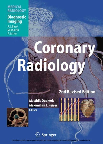
During the past decade, coronary radiology has undergone rapid development. This second edition of the only available monograph on the subject places special emphasis on the role of non-invasive techniques, which can supply information on the condition of the coronary arteries within one simple and short examination. The modalities considered in detail include CT angiography with multidetector and dual-source tomography, 2D and 3D visualization techniques, and MR coronary angiography. Invasive procedures are not neglected, however, and a separate section includes chapters on conventional catheterization, quantitative angiography, and intravascular and quantitative ultrasound. In addition, a section devoted to coronary calcification clearly explains its development and the use of modern techniques in its visualization and quantification. The informative text is supported by a large number of high-quality color images of the coronary and cardiac anatomy.
1;Foreword;5 2;Preface;6 3;Table of contents;8 4;1 Coronary Anatomy;10 4.1;1.1 Introduction;10 4.2;1.2 Physiological and Anatomical Bases;11 4.3;1.3 Normal Coronary Anatomy;14 4.3.1;1.3.1 Left Coronary Anatomy;15 4.3.1.1;1.3.1.1 Left Main Coronary Artery;15 4.3.1.2;1.3.1.2 Left Anterior Descending Coronary Artery;15 4.3.1.3;1.3.1.3 The Circumflex Artery;16 4.3.2;1.3.2 Right Coronary Anatomy;16 4.3.2.1;1.3.2.1 Right Coronary Artery;16 4.3.2.2;1.3.2.2 Determination of Dominance;17 4.4;1.4 Visualization of the Normal Coronary Artery Tree ;17 4.4.1;1.4.1 Anatomy on Catheter Coronary Angiography;17 4.4.2;1.4.2 Anatomy on Multi-detector CT;17 4.5;1.5 Anomalies of the Coronary Artery Tree;27 4.5.1;1.5.1.1 Coronary Artery Fistula;27 4.5.2;1.5.1.2 Anomalous Origin of a Coronary Artery from the Pulmonary Trunk ;28 4.5.3;1.5.1.3 Myocardial Bridging;29 4.6;1.5.2 Vessels Originating from the Contralateral Side, With an Interarterial Course;29 4.6.1;1.5.2.1 Right-Sided Left Coronary Artery;29 4.6.2;1.5.2.2 Left-Sided Right Coronary Artery;29 4.6.3;1.5.2.3 Congenital Coronary Anomalies Not Causing Myocardial Ischemia;32 4.7;References;33 5;2 Invasive Coronary Imaging;34 5.1;2.1 Conventional Catheterisation;34 5.1.1;2.1.1 The Development of Cardiac Catheterisation;34 5.1.1.1;2.1.1.1 Cardiac and Coronary Catheters;35 5.1.1.2;2.1.1.2 Cardiac Catheterisation Laboratories;36 5.1.1.3;2.1.1.3 Radiation Safety and Exposure;36 5.1.1.4;2.1.1.4 Coronary Contrast Injection;37 5.1.1.5;2.1.1.5 Views for Coronary Angiography;37 5.1.1.6;2.1.1.6 Quantifi cation of Coronary Angiography;37 5.1.2;2.1.2 Normal Coronary Anatomy;38 5.1.3;2.1.3 Arterial Dominance;38 5.1.3.1;2.1.3.1 Normal Variants;38 5.1.3.2;2.1.3.2 Muscle Bridge;38 5.1.3.3;2.1.3.3 Coronary Artery Anomalies Presenting in the Adult;39 5.1.4;2.1.4 Coronary Abnormalities Presenting in Infancy and Childhood;39 5.1.4.1;2.1.4.1 Coronary Fistulae;39 5.1.5;2.1.5 Risks of Coronary Angiography;40 5.1.5.1;2.1.5.1 Selection of Patients for Coronary Angiography;40 5.1.5.2;2.1.5.2 Development of Coronary Revascularisation;41 5.1.6;2.1.6 Percutaneous Coronary Intervention;41 5.1.6.1;2.1.6.1 Concomitant Pharmacological Therapy;45 5.1.6.2;2.1.6.2 Long-Term Outcome;46 5.1.6.3;2.1.6.3 Comparison with Bypass Surgery;47 5.1.6.4;2.1.6.4 Percutaneous Coronary Intervention Versus Conservative Therapy;47 5.1.7;2.1.7 Alternative Strategies to the First Line Use of Coronary Angiography;47 5.1.8;References;47 5.2;2.2 Quantitative Coronary Arteriography;50 5.2.1;2.2.1 Introduction;50 5.2.2;2.2.2 Analog or Digital Image Acquisition and Analysis;52 5.2.3;2.2.3 Brief History of Quantitative Coronary Arteriography;52 5.2.4;2.2.4 Basic Principles of Quantitative Coronary Arteriography;53 5.2.4.1;2.2.4.1 Basic Principles of Automated Contour Detection;53 5.2.4.2;2.2.4.2 Calibration Procedure;55 5.2.4.3;2.2.4.3 Coronary Segment Analysis;55 5.2.4.4;2.2.4.4 The Flagging Procedure;56 5.2.4.5;2.2.4.5 Complex Vessel Morphology;58 5.2.4.6;2.2.4.6 Densitometry;59 5.2.4.7;2.2.4.7 Standardized QCA Methodology for Assessing Brachytherapy Trials;60 5.2.4.8;2.2.4.8 Standardized QCA Methodology for Drug-Eluting Stent Analyses;60 5.2.4.8.1;2.2.4.8.1 Definition and Assessment of the Segments Based on Markers or Physical Landmarks ;60 5.2.4.8.2;2.2.4.8.2 Definition and Assessment of the Segments Based on Biologically Relevant Landmarks ;61 5.2.4.8.3;2.2.4.8.3 Definition and Assessment of the "Normal" Segment ;62 5.2.4.8.4;2.2.4.8.4 Definition and Assessment of the Related Parts ;62 5.2.4.8.5;2.2.4.8.5 Data Collection and Reporting for Drug-Eluting Stent Trials;63 5.2.4.9;2.2.4.9 Ostial Analysis;63 5.2.4.10;2.2.4.10 Bifurcation Analysis;65 5.2.5;2.2.5 Guidelines;66 5.2.5.1;2.2.5.1 Guidelines for Catheter Calibration;66 5.2.5.2;2.2.5.2 Guidelines for QCA Acquisition Procedures;67 5.2.6;2.2.6 QCA Validation;68 5.2.6.1;2.2.6.1 Plexiglass Phantom Studies;68 5.2.6.2;2.2.6.2 In Vivo Plexiglass Plugs;69 5.2.6.3;2.2.6.3 Inter- and Intra-observer and Shor
Oudkerk, Matthijs
Reiser, Maximilian F.
Baert, Albert L.
| ISBN | 9783540329848 |
|---|---|
| Artikelnummer | 9783540329848 |
| Medientyp | E-Book - PDF |
| Auflage | 2. Aufl. |
| Copyrightjahr | 2010 |
| Verlag | Springer-Verlag |
| Umfang | 357 Seiten |
| Sprache | Englisch |
| Kopierschutz | Digitales Wasserzeichen |

