Differential Diagnosis in Conventional Radiology
Differential Diagnosis in Conventional Radiology
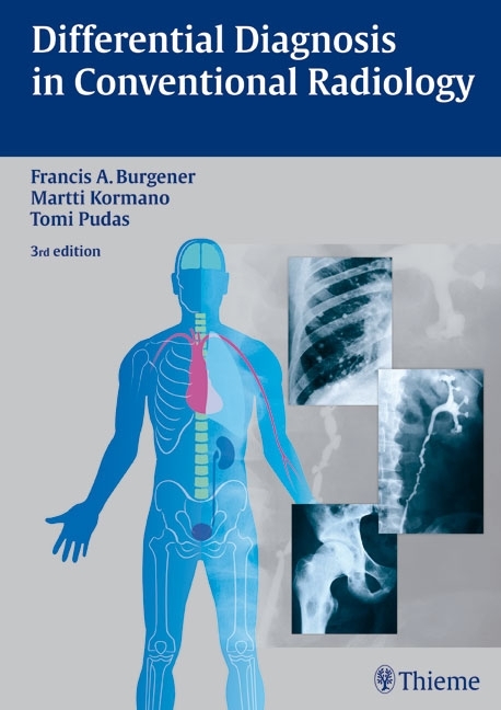
The third updated and revised edition of Differential Diagnosis in Conventional Radiology provides essential information to make conventional x-ray an effective tool in diagnosing disorders affecting the bones and joints and the thoracic and abdominal body segments.
The book is organized according to classifications of radiologic findings rather than disease, enabling the reader to approach diagnosis in a way that reflects the actual clinical situation. Concise and comprehensive tables outline key information on diagnosis and differential diagnosis.
Highlights:
- The unique organization of chapters based on radiologic findings mirrors the situations encountered in daily clinical practice
- Easy-to-reference tables classifying findings, diagnosis, and differential diagnosis and providing important clinical data are perfect for an at-a-glance review
- More than 2000 radiographs and schematic diagrams help to guide the reader toward the most likely diagnoses
The third edition of Differential Diagnosis in Conventional Radiology contains an updated and revised section on radiology of the abdomen combined with the complete text from the recently published books Bone and Joint Disorders and The Chest X-Ray by the same authors.
An exceptional reference work to have on hand, Differential Diagnosis in Conventional Radiology will benefit radiologists and specialists seeking to improve their skills in diagnostic imaging and will also be of great interest to residents preparing for their specialist examinations.
Bone
1 Osteopenia
2 Osteosclerosis
3 Periosteal Reactions
4 Trauma and Fractures
5 Localized Bone Lesions
6 Joint Diseases
7 Joint and Soft-Tissue Calcification
8 Skull
9 Orbits
10 Nasal Fossa and Paranasal Sinuses
11 Jaws and Teeth
12 Spine and Pelvis
13 Clavicles, Ribs, and Sternum
14 Extremities
15 Hands and Feet
Chest
16 Cardiac Enlargement
17 Mediastinal or Hilar Enlargement
18 Pleura and Diaphragm
19 Intrathoracic Calcification
20 Alveolar Infiltrates and Atelectasis
21 Interstitial Lung Disease
22 Pulmonary Edema and Symmetrical Bilateral Infiltrates
23 Pulmonary Nodules and Mass Lesions
24 Pulmonary Cavitary and Cystic Lesions
25 Hyperlucent Lung
Abdomen
26 Abnormal Abdominal Gas Patterns and Dilatation and Motility Disorders in the Gastrointestinal Tract
27 Abdominal Calcifications
28 Abnormal Mucosal Pattern in the Gastrointestinal Tract
29 Narrowing in the Gastrointestinal Tract
30 Filling Defects in the Gastrointestinal Tract
31 Ulcers, Diverticula, and Fistulas in the Gastrointestinal Tract
32 Gallbladder and Bile Duct Abnormalities
33 Abnormal Renal Papillae and Calices
34 Filling Defects in the Urinary Tract
35 Urinary Tract Obstruction and Dilatation
Burgener, Francis A.
Kormano, Martti
Pudas, Tomi
| ISBN | 9783136561034 |
|---|---|
| Artikelnummer | 9783136561034 |
| Medientyp | Buch |
| Auflage | 3rd ed. |
| Copyrightjahr | 2008 |
| Verlag | Thieme, Stuttgart |
| Umfang | 872 Seiten |
| Abbildungen | 1870 Abb. |
| Sprache | Englisch |


