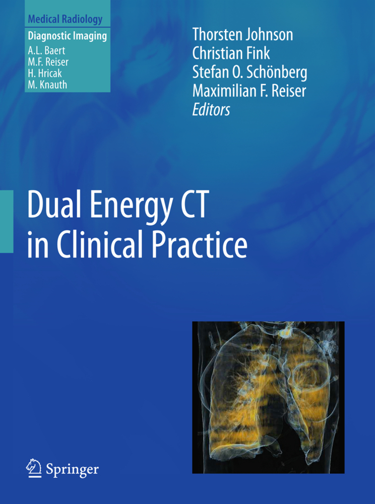
Dual-energy CT is a novel, rapidly emerging imaging technique which offers important new functional and specific information. With implementation of the technology in commercially available scanners, many clinical applications are now feasible.
In this book, physicists and specialists from different CT manufacturers provide an insight into the technological basis of, and the different approaches to, dual-energy CT. Renowned medical scientists in the field explain the pathophysiological and molecular background of the technique, discuss its applications, provide detailed advice on how to obtain optimal results, and offer hints regarding clinical interpretation. The main focus is on the use of dual-energy CT in daily clinical practice, and individual sections are devoted to imaging of the vascular system, the thorax, the abdomen, and the extremities. Evaluations and recommendations are based on personal experience and peer-reviewed literature. Plenty of carefully chosen high-quality images are included to illustrate the clinical benefits of the technique.
In this book, physicists and specialists from different CT manufacturers provide an insight into the technological basis of, and the different approaches to, dual-energy CT. Renowned medical scientists in the field explain the pathophysiological and molecular background of the technique, discuss its applications, provide detailed advice on how to obtain optimal results, and offer hints regarding clinical interpretation. The main focus is on the use of dual-energy CT in daily clinical practice, and individual sections are devoted to imaging of the vascular system, the thorax, the abdomen, and the extremities. Evaluations and recommendations are based on personal experience and peer-reviewed literature. Plenty of carefully chosen high-quality images are included to illustrate the clinical benefits of the technique.
1;Dual Energy CT in Clinical Practice;3 1.1;Copyright Page;4 1.2;Foreword;5 1.3;Preface;6 1.4;Contents;7 1.5;Part I Physical Implementation;9 1.5.1;Physical Background;10 1.5.1.1;1 History;10 1.5.1.2;2 X-ray Spectra;11 1.5.1.3;3 Detector Technology;11 1.5.1.4;4 Tissue Properties;12 1.5.1.5;5 Dual-Source CT;12 1.5.1.6;6 Rapid Voltage Switching;13 1.5.1.7;7 Layer Detector;14 1.5.1.8;8 Sequential Acquisition;14 1.5.1.9;9 Radiation Exposure;15 1.5.1.10;10 Clinical Applications;15 1.5.1.11;11 Summary;15 1.5.1.12;References;15 1.5.2;Dual Source CT;17 1.5.2.1;1 Dual Source Dual Energy Scanning;18 1.5.2.1.1;1.1 Introduction;18 1.5.2.1.2;1.2 Scanner Design;18 1.5.2.1.2.1;1.2.1 SOMATOM Definition;18 1.5.2.1.2.2;1.2.2 SOMATOM Definition Flash;19 1.5.2.1.3;1.3 Data Acquisition;20 1.5.2.2;2 Image-Based DECT Analysis;22 1.5.2.2.1;2.1 Introduction;22 1.5.2.2.2;2.2 The Thin Absorber Case;22 1.5.2.2.3;2.3 Mixed Image, Monoenergetic Image, Dual Energy Index;23 1.5.2.2.4;2.4 Material Decomposition Analysis;24 1.5.2.2.5;2.5 Material Labeling and Highlighting;24 1.5.2.3;3 Outlook;25 1.5.2.4;References;25 1.5.3;Dual Layer CT;27 1.5.3.1;1 Introduction;28 1.5.3.2;2 Physical Background;28 1.5.3.3;3 System Design;30 1.5.3.4;4 Image Reconstruction;30 1.5.3.4.1;4.1 Image Reconstruction and Energy Map;30 1.5.3.4.2;4.2 Material Separation and Noise;31 1.5.3.4.3;4.3 Polychromatic Correction;33 1.5.3.5;5 Post-processing;35 1.5.3.5.1;5.1 The Vectorial Separation;35 1.5.3.5.2;5.2 The Probability Separation;36 1.5.3.5.3;5.3 3D Rendering;36 1.5.3.6;References;40 1.5.4;Gemstone Detector: Dual Energy Imaging via Fast kVp Switching;41 1.5.4.1;1 Physical Background;41 1.5.4.2;2 System Design;42 1.5.4.3;3 Image Reconstruction;43 1.5.4.4;4 Projection-Based Material Decomposition;43 1.5.4.5;5 Post Processing;44 1.5.4.5.1;5.1 Noise Suppression;44 1.5.4.5.2;5.2 GSI Viewer;44 1.5.4.6;6 Future Research/Applications;45 1.5.4.7;References;47 1.5.5;Dual-Energy Algorithms and Postprocessing Techniques;48 1.5.5.1;1 Introduction;48 1.5.5.2;2 How to Process;49 1.5.5.2.1;2.1 Projection Space Approach;49 1.5.5.2.2;2.2 Image Space Approach;50 1.5.5.3;3 Type of DE Image;51 1.5.5.3.1;3.1 Blended Image;51 1.5.5.3.2;3.2 Material-Selective Image;52 1.5.5.3.3;3.3 Energy-Selective Image;54 1.5.5.4;4 Determinants of IQ/Dose;54 1.5.5.5;References;56 1.6;Part II Vascular System;57 1.6.1;Head and Neck;58 1.6.1.1;1 Clinical Background;58 1.6.1.2;2 Scan Protocol;59 1.6.1.3;3 Contrast Material Injection;61 1.6.1.4;4 Postprocessing;61 1.6.1.5;5 Diagnostic Evaluation;61 1.6.1.5.1;5.1 Stroke CT and Stenosis Evaluation;61 1.6.1.5.2;5.2 Virtual Unenhanced Images and Brain Hemorrhage;61 1.6.1.5.3;5.3 Intracranial Aneurysm, Angiomas, and Fistulas;62 1.6.1.6;6 Scientific Evidence;63 1.6.1.7;References;63 1.6.2;Aorta;64 1.6.2.1;1 Clinical Background;64 1.6.2.2;2 Physical Background;65 1.6.2.3;3 Scan Protocol;66 1.6.2.4;4 Contrast Material Injection;66 1.6.2.5;5 Postprocessing;67 1.6.2.6;6 Diagnostic Evaluation;67 1.6.2.7;7 Scientific Evidence;68 1.6.2.8;References;68 1.6.3;Peripheral Arteries;70 1.6.3.1;1 Clinical Background;70 1.6.3.2;2 Physical Background;71 1.6.3.3;3 Scan Protocol;71 1.6.3.4;4 Contrast Material Injection;72 1.6.3.5;5 Postprocessing;72 1.6.3.6;6 Diagnostic Evaluation;72 1.6.3.7;7 Scientific Evidence;72 1.6.3.8;References;74 1.6.4;Plaque Differentiation;76 1.6.4.1;1 Introduction;76 1.6.4.2;2 Rationale of Dual-Energy CT;77 1.6.4.2.1;2.1 Original Intensity Space;78 1.6.4.2.2;2.2 Dual-Energy CT Imaging of Plaques;78 1.6.4.3;3 Potential Future Directions for Dual-Energy CT Plaque Imaging;79 1.6.4.4;4 Conclusion;81 1.6.4.5;References;81 1.7;Part III Thoracic Imaging;83 1.7.1;Lung Perfusion;84 1.7.1.1;1 Clinical Background;85 1.7.1.2;2 Physical Background;85 1.7.1.3;3 Scan Protocol;86 1.7.1.4;4 Contrast Material Injection;87 1.7.1.5;5 Postprocessing;87 1.7.1.6;6 Diagnostic Evaluation;88 1.7.1.7;7 Scientific Evidence;88 1.7.1.8;References;91 1.7.2;Lung Ventilation;92 1.7.2.1;1 Background: Basi
Johnson, Thorsten
Fink, Christian
Schönberg, Stefan O.
Reiser, Maximilian F
| ISBN | 9783642017407 |
|---|---|
| Artikelnummer | 9783642017407 |
| Medientyp | E-Book - PDF |
| Auflage | 2. Aufl. |
| Copyrightjahr | 2011 |
| Verlag | Springer-Verlag |
| Umfang | 216 Seiten |
| Kopierschutz | Digitales Wasserzeichen |

