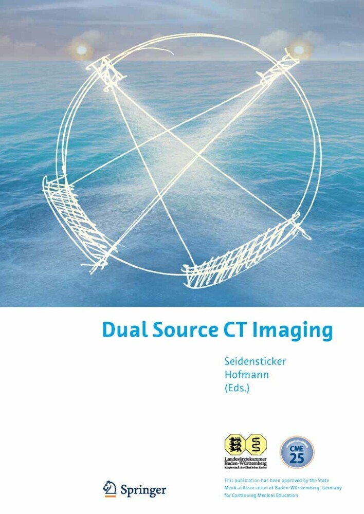
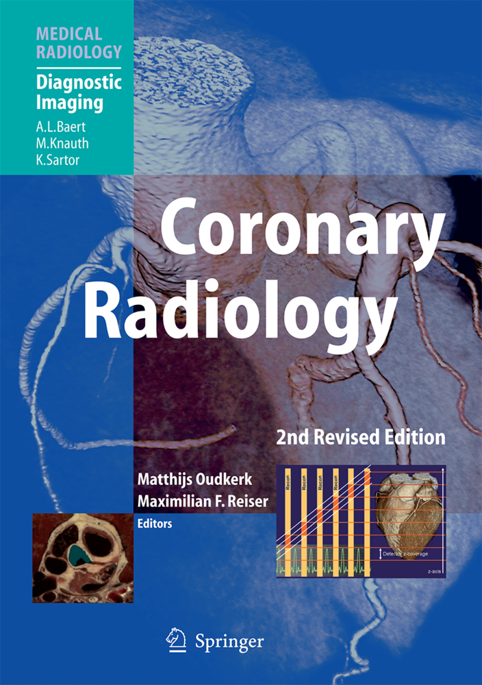
The introduction of Dual Source Computed Tomography (DSCT) in 2005 was an evolutionary leap in the field of CT imaging. Two x-ray sources operated simultaneously enable heart-rate independent temporal resolution and routine spiral dual energy imaging. The precise delivery of contrast media is a critical part of the contrast-enhanced CT procedure.
This book provides an introduction to DSCT technology and to the basics of contrast media administration followed by 25 in-depth clinical scan and contrast media injection protocols. All were developed in consensus by selected physicians on the Dual Source CT Expert Panel. Each protocol is complemented by individual considerations, tricks and pitfalls, and by clinical examples from several of the world's best radiologists and cardiologists.
This extensive CME-accredited manual is intended to help readers to achieve consistently high image quality, optimal patient care, and a solid starting point for the development of their own unique protocols.
1;Content;52;Dear Colleagues;93;The Dual Source CT Expert Panel;114;Authors;155;Dual Source CT Technology;216;Iodinated Contrast Media: New Perspectives with Dual Source CT;377;Cardiac: Coronary CT Angiography;538;Case 1 Ruling out Coronary Stenosis in Acute Chest Pain;589;Case 2 Diagnosis of a Coronary Occlusion in a Patient with Atypical Chest Pain;6010;Cardiac: Coronary Stents;6311;Case 1 Stent in LAD;6812;Case 2 Stent Restenosis;7013;Cardiac: Coronary CTA in Obese Patients;7314;DSCT in a Morbidly Obese Woman;7815;Case 1 DSCT in a Morbidly Obese Woman Presenting with Dyspnea on Exertion;7816;Case 2 DSCT in a Morbidly Obese Woman Presenting with Acute Atypical Chest Pain;8017;Cardiac: Valvular Function;8318;Case 1 Bicuspid Aortic Valve;8819;Case 2 Mitral Regurgitation;9020;Cardiac: Morphology;9321;Case 1 Complex Congenital Heart Disease, s / p Repair;9822;Case 2 Anterior MI, s / p CABG;10023;Cardiac: Left / Right Ventricular Function;10324;Case 1 Dual Source CT Preceding Cardiac Resynchronization Therapy (CRT);10825;Case 2 Dual Source CT in Left-Ventricular Hypertrophy [A] & in ARVCM [B];11026;Cardiac: Atrial Fibrillation / Arrhythmia;11327;Case 1 Patient with Atrial Fibrillation and Atypical Chest Pain;11828;Case 2 Patient with Atrial Fibrillation and Chronic Stable Angina Pectoris;12029;Cardiac: Bypass Grafts;12330;Case 1 Thrombosed Venous Bypass Graft Pseudoaneurysm Following Coil Embolization;12831;Case 2 Quadruple, Patent Coronary Artery Bypass Grafts;13032;Vascular: Extended Chest Pain Protocol;13333;Case 1 Ruling Out of Cardiovascular Causes in Acute Unclear Chest Pain;13834;Case 2 50-Year-Old Male with Unclear Chest Pain;14035;Vascular: Pulmonary Veins;14336;Case 1 Minor Pulmonary Vein Stenosis Post RFCA L;14837;Case 2 Significant Vein Stenosis;15038;Vascular: Aortic Runoff, Abdominal CTA;15339;Case 1 Follow-Up After Transarterial Embolization of a Liver Metastasis;15840;Case 2 Assessment of Peripheral Artery Disease;16041;Vascular: Renal CTA;16342;Case 1 Renal Artery Dissection;16843;Case 2 Living Renal Donor;17044;Vascular: Peripheral Runoff;17345;Case 1 Suspicion of Peripheral Occlusive Disease;17846;Case 2 Suspicion of Femoral Artery Stenosis;18047;Vascular: Brain Perfusion;18348;Case 1 PBV Delineates the Whole Volume of Cerebral Infarction in Hyperacute Phase;18849;Case 2 PBV Shows Small Infarction Missed in Perfusion CT;19050;Body: Obese Mode;19351;Case 1 Aortic Dissection in 183 kg Male;19852;Case 2 Abdominal Abscess in 206 kg Male;20053;Dual Energy: CTA of Head and Neck;20554;Case 1 Multi-segmental Stenosis of all Cervical Vessels and Intracranial Aneurysm;21055;Case 2 Stenosis of Internal Carotid Artery;21256;Dual Energy: CTA Aorta;21557;Case 1 Abdominal Aortic Aneurysm and Interventional Stent Placement;22058;Prosthesis Infection After Surgical;22259;Case 2 Prosthesis Infection After Surgical Abdominal Aortic Prosthesis;22260;Dual Energy: CTA Runoff;22561;Case 1 Diabetic Female with Bilateral Calf Pain;23062;Case 2 Elderly Hypertensive Male with Severe Leg Pain;23263;Dual Energy: CTA Lung Perfusion (PE);23564;Case 1 Acute Pulmonary Embolism;24065;Case 2 Pulmonary Hypertension Due to Chronic Recurrent Pulmonary Embolism;24266;Dual Energy: Virtual Non-Contrast;24567;Case 1 Scanning of a Small Polyp in Gallbladder;25068;Case 2 Exclusion of Urolithiasis in the Presence of Contrast Media;25269;Dual Energy: Characterization of Kidney Stone Composition;25570;Case 1 Ex Vivo Validation of Algorithm Accuracy;26071;Case 2 In Vivo Characterization of Large Renal Stone;26272;Dual Energy: Urography;26573;Virtual Non-Contrast CT;27074;Case 1 Virtual Non-Contrast CT for Identification of Stones from Contrast Enhanced CT;27075;Case 2 Dual Energy CT for the Differentiation of Renal Cyst vs. Mass;27276;Dual Energy: Vascular Plaque Removal / Detection;27577;Case 1 Calcification of the Carotid Artery;28078;Case 2 Calcification of the Renal Artery;2827
Seidensticker, Peter R.
Hofmann, Lars K.
| ISBN | 9783540776024 |
|---|---|
| Artikelnummer | 9783540776024 |
| Medientyp | E-Book - PDF |
| Auflage | 2. Aufl. |
| Copyrightjahr | 2008 |
| Verlag | Springer-Verlag |
| Umfang | 290 Seiten |
| Sprache | Englisch |
| Kopierschutz | Adobe DRM |

