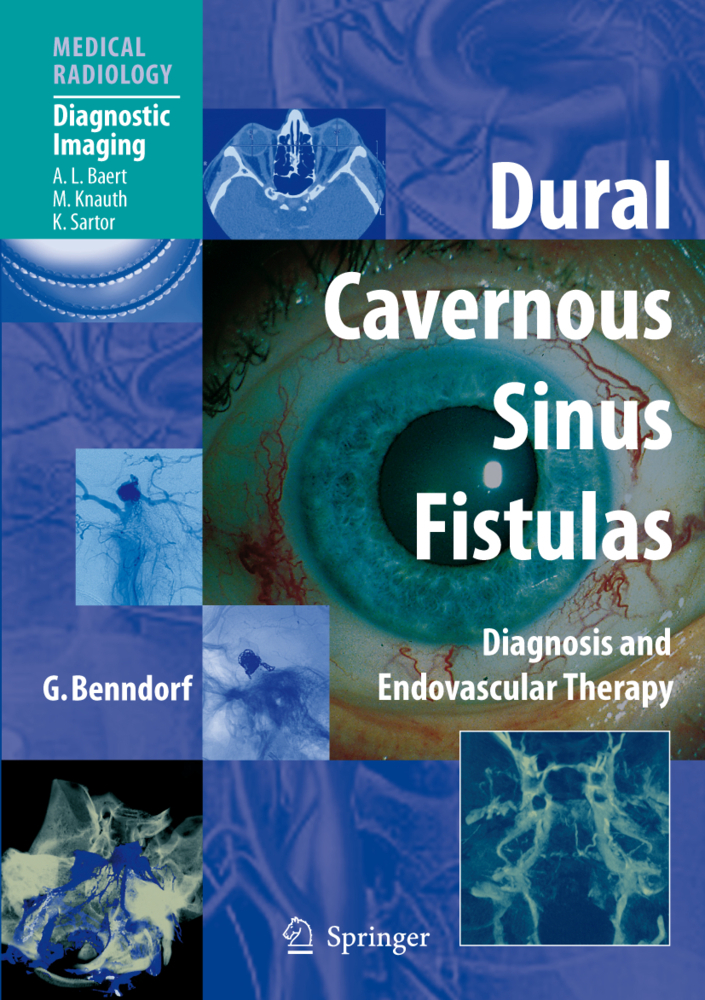
Dural Cavernous Sinus Fistulas
Diagnosis and Endovascular Therapy
Dural cavernous sinus fistulas (DCSFs) are benign vascular diseases consisting in an arteriovenous shunt at the cavernous sinus that if misdiagnosed can lead to potentially serious ophthalmologic complications. This volume provides a complete guide to the diagnosis and minimal invasive treatment of DCSFs. After sections on anatomy and classification, etiology and pathogenesis of DCSFs, the symptomatology of the disease is described in detail. The role of modern imaging techniques in the diagnosis of DCSFs is then addressed. Digital subtraction angiography (DSA) remains the gold standard for clinical decision-making; here, full consideration is given to both, conventional 2D DSA and rotational 3D angiography. Recent technological advances in this field such as Dual Volume (DV) imaging and angiographic computed tomography (ACT) are considered as well. Due attention is further paid to the use of computed tomography, magnetic resonance imaging and ultrasound. Finally, the therapeutic management of DCSFs with emphasis on various transvenous occlusion techniques are discussed in depth. This well-illustrated volume will be invaluable to all who may encounter DCSF in their clinical practice.
1;Foreword;6 2;Preface;7 3;Table of Contents;9 4;Glossary;11 5;1 Introduction;13 5.1;References;14 6;2 Historical Considerations;15 6.1;2.1 Arteriovenous Fistula and Pulsating Exophthalmos;15 6.2;2.2 Angiography;18 6.3;2.3 Therapeutic Measures;21 6.4;2.4 Embolization;22 6.5;References;23 7;3 Anatomy of the Cavernous Sinus and Related Structures;26 7.1;3.1 Osseous Anatomy;26 7.1.1;3.1.1 Orbit;29 7.2;3.2 Anatomy of the Dura Mater and the Cranial Nerves;31 7.2.1;3.2.1 Autonomic Nervous System;32 7.3;3.3 Vascular Anatomy;32 7.3.1;3.3.1 Arterial Anatomy;32 7.3.1.1;3.3.1.1 Internal Carotid Artery;32 7.3.1.1.1;3.3.1.1.1 Branches of the ICA Branches of the Cavernous Segment;34 7.3.1.1.1.1;Meningohypophyseal Trunk (MHT);34 7.3.1.1.1.2;The inferolateral trunk (ILT);35 7.3.1.1.1.3;Ophthalmic Artery;37 7.3.1.1.1.4;Ethmoidal Arteries;39 7.3.1.2;3.3.1.2 External Carotid Artery;40 7.3.1.2.1;3.3.1.2.1 Ascending Pharyngeal Artery;40 7.3.1.2.2;3.3.1.2.2 Internal Maxillary Artery;40 7.3.1.2.3;3.3.1.2.3 Middle Meningeal Artery;41 7.3.1.2.4;3.3.1.2.4 Accessory Meningeal Artery;41 7.3.2;3.3.2 Venous Anatomy;42 7.3.2.1;3.3.2.1 The Cavernous Sinus, Receptaculum, Sinus Caroticus (Rektorzik), Con. uens Sinuum Anterius, Sinus Spheno-Parietale (Cruveilhier), Cavernous Plexus, Lateral Sellar Compartment;42 7.3.2.1.1;3.3.2.1.1 Embryology;42 7.3.2.1.2;3.3.2.1.2 Anatomy and Topography;42 7.3.2.2;3.3.2.2 Tributaries of the Cavernous Sinus (Afferent Veins);47 7.3.2.2.1;Orbital Veins;47 7.3.2.2.2;Superior Ophthalmic Vein;47 7.3.2.2.3;Inferior Ophthalmic Vein;48 7.3.2.2.4;Central Retinal Vein (No Direct CS Tributary);49 7.3.2.2.5;Superficial Middle Cerebral Vein, Sylvian Vein;49 7.3.2.2.6;Uncal Vein, Uncinate Vein;50 7.3.2.2.7;Sphenoparietal sinus (Breschet), Sinus alae parvae, Sinus sphenoidales superior (Sir C. Bell);50 7.3.2.2.8;Intercavernous Sinus, Sinus intercavernosus, Sinus circularis (Ridley), Sinus ellipticus, Sinus coronarius, Sinus clinoideus (Sir C. Bell), Sinus transversus sellae equinae (Haller);50 7.3.2.2.9;Meningeal Veins;51 7.3.2.2.10;Veins of the Foramen Rotundum, Emissary Vein;51 7.3.2.3;3.3.2.3 Drainage of the Cavernous Sinus (Efferent Veins);51 7.3.2.3.1;Superior Petrosal Sinus, Sinus petrobasilaris (Langer), Sinus tentorii lateralis (Weber), Sinus petrosus super. cialis;51 7.3.2.3.2;Inferior Petrosal Sinus, Sinus petrosus profundus, Sinus petro-occipitalis superior (Trolard);51 7.3.2.3.3;Venous Plexus of the Hypoglossal Canal, Anterior Condylar Vein;53 7.3.2.3.4;Posterior Condylar Vein;53 7.3.2.3.5;Lateral Condylar Vein;53 7.3.2.3.6;Inferior Petroclival Vein;53 7.3.2.3.7;Petro-occipital Sinus, Sinus petro-occipitalis inferior, petro-occipital vein (Padget);53 7.3.2.3.8;Transverse Occipital Sinus (Doyen);53 7.3.2.3.9;Basilar Plexus (Virchow);53 7.3.2.3.10;Marginal Sinus;53 7.3.2.3.11;Internal Carotid Artery Venous Plexus, Sinus Venous Caroticus (Haike), Carotid Sinus, Pericarotid Plexus;54 7.3.2.3.12;Foramen Ovale Plexus (Trigeminal Sinus), Sphenoid Emissary, "Rete" of the Foramen Ovale;54 7.3.2.3.13;Vein of the Sphenoid Foramen (Foramen Venosum, Foramen of Vesalius);54 7.3.2.3.14;Foramen Lacerum Plexus;54 7.3.2.3.15;Pterygoid Plexus;54 7.3.2.4;3.3.2.4 Other Veins of Importance for the CS Drainage or for Transvenous Access to the CS;55 7.3.2.4.1;Facial Vein;55 7.3.2.4.2;Frontal Vein;55 7.3.2.4.3;Angular Vein;55 7.3.2.4.4;Middle Temporal Vein;55 7.3.2.4.5;Internal Jugular Vein;55 7.3.2.4.6;The External Jugular Vein;56 7.3.2.4.7;Vertebral Vein, Vertebral Artery Venous Plexus;56 7.3.2.4.8;Deep Cervical Vein;56 7.3.2.4.9;Anterior Condylar Con. uent (Con. uens Condyloideum Anterius, Trolard 1868);57 7.4;References;57 8;4 Classification of Cavernous Sinus Fistulas (CSFs) and Dural Arteriovenous Fistulas (DAVFs);62 8.1;Introduction;62 8.2;4.1 Anatomic Classification;62 8.2.1;4.1.1 Dural Arteriovenous Fistulas (DAVFs);62 8.2.2;4.1.2 Cavernous Sinus Fistulas (CSFs);65 8.3;4.2 Etiologic Classification;71 8.4;4.3 Hemodynamic Classification;71 8.5;Refe
Benndorf, Goetz
| ISBN | 9783540688891 |
|---|---|
| Artikelnummer | 9783540688891 |
| Medientyp | E-Book - PDF |
| Auflage | 2. Aufl. |
| Copyrightjahr | 2010 |
| Verlag | Springer-Verlag |
| Umfang | 320 Seiten |
| Kopierschutz | Digitales Wasserzeichen |

