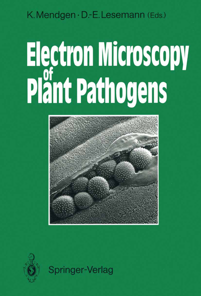
Electron Microscopy of Plant Pathogens
Electron Microscopy of Plant Pathogens
Plants, fungi, and viruses were among the first biological objects studied with an electron microscope. One of the two first instruments built by Siemens was used by Helmut Ruska, a brother of Ernst Ruska, the pioneer in constructing electron microscopes. H. Ruska published numerous papers on different biological objects in 1939. In one of these, the pictures by G. A. Kausche, E. Pfankuch, and H. Ruska of tobacco mosaic virus opened a new age in microscopy. The main problem was then as it still is today, to obtain an appropriate preparation of the specimen for observation in the electron microscope. Beam damage and specimen thickness were the first obstacles to be met. L. Marton in Brussels not only built his own instrument, but also made considerable progress in specimen preparation by introducing the impregnation of samples with heavy metals to obtain useful contrast. His pictures of the bird nest orchid root impregnated with osmium were revolutionary when published in 1934. It is not the place here to recall the different techniques which were developed in the subsequent years to attain the modern knowledge on the fine structure of plant cells and of different plant pathogens. The tremendous progress obtained with tobacco mosaic virus is reflected in the chapter by M. Wurtz on the fine structure of viruses in this Volume. New cytochemical and immunological techniques considerably surpass the morphological information obtained from the pathogens, especially at the host-parasite interface.
3 High Pressure Freezing of Rust Infected Plant Leaves
4 Cytochemistry of Fungal Surfaces: Carbohydrate Containing Molecules
5 Analytical Electron Microscopy in Plant Pathology: X-Ray Microanalysis and Energy Loss Spectroscopy
6 The Fine Structure of Virus Particles
7 Immunoelectron Microscopy for Virus Identification
8 Cytochemistry of Virus-Infected Plant Cells
9 Immunolabeling of Viral Antigens in Infected Cells
10 Mechanisms of Plant Virus Transmission by Homopteran Insects
11 Specific Cytological Alterations in Virus-Infected Plant Cells
12 Structure, Cellular Location, and Cytopathology of Viroids
13 Immunological Detection and Localization of Mycoplasma-Like Organisms (MLOs) in Plants and Insects by Light and Electron Microscopy
14 Interactions Between Pseudomonas and Phaseolus vulgaris
15 The Fate of Peripheral Vesicles in Zoospores of Phytophthora cinnamomi During Infection of Plants
16 Hemibiotrophy in Colletotrichum lindemuthianum
17 Extracellular Materials of Fungal Structures: Their Significance at Prepenetration Stages of Infection
18 Rust Haustoria
19 Infection by Magnaporthe: An In Vitro Analysis
20 Mycorrhizal and Pathogenic Fungi: Do They Share Any Features?
21 Haustoria-Like Structures and Hydrophobic Cell Wall Surface Layers in Lichens
22 Ultrastructure of Nematode-Plant Interactions
23 Infection of Plants by Flagellate Protozoa (Phytomonas SPP., Trypanosomatidae)
24 Influence of Fungicides on Fungal Fine Structure.
1 Preservation of Cell Ultrastructure by Freeze-Substitution
2 Low-Temperature Scanning Electron Microscopy of Fungi and Fungus-Plant Interactions3 High Pressure Freezing of Rust Infected Plant Leaves
4 Cytochemistry of Fungal Surfaces: Carbohydrate Containing Molecules
5 Analytical Electron Microscopy in Plant Pathology: X-Ray Microanalysis and Energy Loss Spectroscopy
6 The Fine Structure of Virus Particles
7 Immunoelectron Microscopy for Virus Identification
8 Cytochemistry of Virus-Infected Plant Cells
9 Immunolabeling of Viral Antigens in Infected Cells
10 Mechanisms of Plant Virus Transmission by Homopteran Insects
11 Specific Cytological Alterations in Virus-Infected Plant Cells
12 Structure, Cellular Location, and Cytopathology of Viroids
13 Immunological Detection and Localization of Mycoplasma-Like Organisms (MLOs) in Plants and Insects by Light and Electron Microscopy
14 Interactions Between Pseudomonas and Phaseolus vulgaris
15 The Fate of Peripheral Vesicles in Zoospores of Phytophthora cinnamomi During Infection of Plants
16 Hemibiotrophy in Colletotrichum lindemuthianum
17 Extracellular Materials of Fungal Structures: Their Significance at Prepenetration Stages of Infection
18 Rust Haustoria
19 Infection by Magnaporthe: An In Vitro Analysis
20 Mycorrhizal and Pathogenic Fungi: Do They Share Any Features?
21 Haustoria-Like Structures and Hydrophobic Cell Wall Surface Layers in Lichens
22 Ultrastructure of Nematode-Plant Interactions
23 Infection of Plants by Flagellate Protozoa (Phytomonas SPP., Trypanosomatidae)
24 Influence of Fungicides on Fungal Fine Structure.
Mendgen, Kurt
Lesemann, Dietrich-Eckhardt
| ISBN | 978-3-642-75820-1 |
|---|---|
| Artikelnummer | 9783642758201 |
| Medientyp | Buch |
| Copyrightjahr | 2011 |
| Verlag | Springer, Berlin |
| Umfang | XV, 336 Seiten |
| Abbildungen | XV, 336 p. |
| Sprache | Englisch |

