The technology of angiographic systems has been improved tremendously just within the past few years. This has allowed greatly increased levels of image resolution for both fluorescein and indocyanine green angiography.
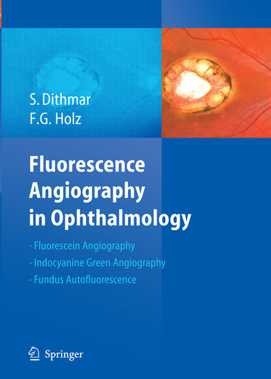
This new atlas by Dithmar and Holz covers the basic principles of the new methods for fluorescein- and indocyanine green-angiography, as well as the high resolution imaging of fundus autofluorescence.
The angiographic signs of retinal and choroidal diseases are illustrated with images taken from a series of clinically relevant case examples that specifically illustrate the advantages of higher image resolution for the study of common retinochoroidal disorders. In so doing, this atlas offers an all-encompassing survey of the many angiographic signs in these disorders and their differential diagnoses. Clinicians in all subspecialties of ophthalmology can profit from a better understanding of these pathophysiological phenomena.
1;Preface;5 2;Foreword;7 3;Table of Contents;9 4;The physical and chemical fundamentals of fluorescence angiography;11 4.1;Fluorescence;12 4.2;Fluorescein;12 4.3;Indocyanine Green;13 5;The Technical Fundamentals of Fluorescence Angiography;15 5.1;The basic design of a scanning laser ophthalmoscope;16 5.2;Light Sources;17 5.3;Lasers for Fluorescein Angiography;17 5.4;Lasers for red- free fundus imaging;18 5.5;Lasers for Indocyanine green ( ICG) angiography;19 5.6;Lasers for reflective infrared imaging;19 5.7;The Fundamentals of Optical Imaging The confocal principle;19 5.8;Resolution in depth;20 5.9;Resolution in breadth;20 5.10;The Heidelberg Retina Angiograph 2;20 5.11;HRA2- Parameters for basic imaging modes;21 5.12;Simultaneous mode;21 5.13;Composite mode;22 5.14;Fixation assistance;22 5.15;Wide angle objective;22 5.16;ART module;22 5.17;Examination of the anterior segment;22 5.18;Stereoscopic imaging;22 5.19;Components of the analytical software;23 6;Normal Fluorescence Angiography and General Pathological Fluorescence Phenomena;25 6.1;Normal Fluorescence Angiography and general pathological fluorescence phenomena;26 6.2;Choroid;26 6.3;Cilioretinal vessels;26 6.4;Optic disc;26 6.5;Retinal vessels;26 6.6;Macula;26 6.7;Sclera;27 6.8;Iris;27 6.9;Normal ICG- Angiography;27 6.10;Pathological fluorescence phenomena;27 6.11;Hyperfluorescence;27 6.12;Hypofluorescence;28 7;Fundus Autofluorescence;41 7.1;Introduction;42 7.2;Scanning laser ophthalmoscopy and modified fundus photography;42 7.3;Methods Fundamentals;42 7.4;The operational sequence of a fundus examination with the cSLO;42 7.5;Lipofuscin in the retinal pigment epithelium;43 7.6;Normal fundus autofluorescence;43 7.7;Fundus autofluorescence - typical findings;43 7.8;Lipofuscin Melanin;45 8;Macular Disorders;65 8.1;Age- Related Macular Degeneration ( AMD);66 8.2;Drusen;66 8.3;Irregular Pigmentation of the Retinal Pigment Epithelium ( RPE);72 8.4;Geographic atrophy of the RPE;72 8.5;Choroidal Neovascularization ( CNV);74 8.6;Classic Choroidal Neovascularization;74 8.7;Occult Choroidal Neovascularization;79 8.8;Mixed Forms;84 8.9;Localization of Choroidal Neovascularization;84 8.10;Serous Detachment of the Retinal Pigment Epithelium;86 8.11;Breaks in the Retinal Pigment Epithelium ( RPE);92 8.12;Idiopathic Polypoid Choroidal Vasculopathy;94 8.13;Retinal Angiomatous Proliferation ( RAP);96 8.14;Disciform Scarring;98 8.15;Fluorescein Angiographic Phenomena Following Therapy;100 8.16;Laser Coagulation;100 8.17;Photodynamic Therapy ( PDT);100 8.18;Anti VEGF Therapy;100 8.19;Cystoid Macular Edema;112 8.20;Hereditary Macular Disorders Stargardt's Disease;114 8.21;Fundus Flavimaculatus;114 8.22;Best's Disease ( Vitelliform Macular dystrophy);116 8.23;Pattern Dystrophies of the Retinal Pigment Epithelium ( RPE);118 8.24;Congenital X- Chromosome Retinoschisis;120 8.25;Adult Vitelliform Macular Degeneration ( AVMD);122 8.26;The Macular Degeneration of Myopia;124 8.27;Angioid Streaks;126 8.28;Central Serous Chorioretinopathy ( CSC);128 8.29;Chronic Idiopathic Central Serous Chorioretinopathy ( CICSC);130 8.30;Idiopathic Juxtafoveal Teleangiectasias;132 8.31;Epiretinal Gliosis;134 8.32;Macular Holes;136 8.33;Chloroquine Maculopathy;138 9;Retinal Vascular Disease;141 9.1;Diabetic Retinopathy Nonproliferative Diabetic Retinopathy;142 9.2;Proliferative Diabetic Retinopathy;144 9.3;Hypertensive Retinopathy;146 9.4;Retinal Arterial Occlusions Occlusion of the Central Retinal Artery;148 9.5;Branch Retinal Artery Occlusion;148 9.6;Retinal Vein Occlusions;150 9.7;Central Retinal Vein Occlusion ( CRVO);150 9.8;Tributary Retinal Vein Occlusion;154 9.9;Retinal Macroaneurysms;156 9.10;Coats' Disease;158 9.11;Retinal Capillary Hemangioma;160 9.12;Cavernous Hemangioma of the Retina;162 9.13;Vascular Tortuosity;164 10;Inflammatory Retinal/Choroidal Disease;167 10.1;Toxoplasmic Retinochoroiditis;168 10.2;Multifocal Choroiditis;170 10.3;Acute Posterior Multifocal Placoid Pigment Epitheliopathy ( APMPPE);1
| ISBN | 9783540794011 |
|---|---|
| Artikelnummer | 9783540794011 |
| Medientyp | E-Book - PDF |
| Copyrightjahr | 2008 |
| Verlag | Springer-Verlag |
| Umfang | 224 Seiten |
| Sprache | Englisch |
| Kopierschutz | Digitales Wasserzeichen |

