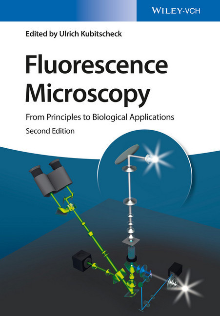
Fluorescence Microscopy
From Principles to Biological Applications
While there are many publications on the topic written by experts for experts, this text is specifically designed to allow advanced students and researchers with no background in physics to comprehend novel fluorescence microscopy techniques.
This second edition features new chapters and a subsequent focus on super-resolution and single-molecule microscopy as well as an expanded introduction. Each chapter is written by a renowned expert in the field, and has been thoroughly revised to reflect the developments in recent years.
Ulrich Kubitscheck is the department head of Biophysical Chemistry at the Institute of Physical and Theoretical Chemistry at the University of Bonn, Germany. Having obtained his academic degrees from the University of Bremen, Germany, he spent his career working at The Weizmann Institute of Science and the Institute of Medical Physics and Biophysics of the University of Munster, Germany, before taking up his present appointment at the University of Bonn. Professor Kubitscheck develops single molecule and light sheet imaging techniques, has authored over 80 scientific publications and has extensive experience in teaching courses on physical chemistry, biophysics and quantitative microscopy.
1;Cover;1 2;Title Page;5 3;Copyright;6 4;Contents;7 5;List of Contributors;17 6;Preface;21 7;Chapter 1 Introduction to Optics;25 7.1;1.1 A Short History of Theories about Light;25 7.2;1.2 Properties of Light Waves;26 7.2.1;1.2.1 An Experiment on Interference;26 7.2.2;1.2.2 Physical Description of Light Waves;27 7.3;1.3 Four Effects of Interference;31 7.3.1;1.3.1 Diffraction;31 7.3.2;1.3.2 The Refractive Index;33 7.3.3;1.3.3 Refraction;33 7.3.4;1.3.4 Reflection;34 7.3.5;1.3.5 Light Waves and Light Rays;35 7.4;1.4 Optical Elements;37 7.4.1;1.4.1 Lenses;37 7.4.2;1.4.2 Metallic Mirrors;39 7.4.3;1.4.3 Dielectric Mirrors;40 7.4.4;1.4.4 Filters;41 7.4.5;1.4.5 Chromatic Reflectors;42 7.5;1.5 Optical Aberrations;44 7.6;References;46 8;Chapter 2 Principles of Light Microscopy;47 8.1;2.1 Introduction;47 8.2;2.2 Construction of Light Microscopes;47 8.2.1;2.2.1 Components of Light Microscopes;47 8.2.2;2.2.2 Imaging Path;48 8.2.3;2.2.3 Magnification;50 8.2.4;2.2.4 Angular and Numerical Aperture;51 8.2.5;2.2.5 Field of View;52 8.2.6;2.2.6 Illumination Beam Path;52 8.2.6.1;2.2.6.1 Critical and Köhler Illumination;52 8.2.6.2;2.2.6.2 Bright-Field and Epi-Illumination;55 8.3;2.3 Wave Optics and Resolution;56 8.3.1;2.3.1 Wave Optical Description of the Imaging Process;57 8.3.2;2.3.2 The Airy Pattern;61 8.3.3;2.3.3 Point Spread Function and Optical Transfer Function;64 8.3.4;2.3.4 Lateral and Axial Resolution;65 8.3.4.1;2.3.4.1 Lateral Resolution Using Incoherent Light Sources;65 8.3.4.2;2.3.4.2 Lateral Resolution of Coherent Light Sources;67 8.3.4.3;2.3.4.3 Axial Resolution;69 8.3.5;2.3.5 Magnification and Resolution;72 8.3.6;2.3.6 Depth of Field and Depth of Focus;73 8.3.7;2.3.7 Over- and Undersampling;74 8.4;2.4 Apertures, Pupils, and Telecentricity;74 8.5;2.5 Microscope Objectives;77 8.5.1;2.5.1 Objective Lens Design;77 8.5.2;2.5.2 Light Collection Efficiency and Image Brightness;81 8.5.3;2.5.3 Objective Lens Classes;85 8.5.4;2.5.4 Immersion Media;86 8.5.5;2.5.5 Special Applications;89 8.6;2.6 Contrast;91 8.6.1;2.6.1 Dark Field;92 8.6.2;2.6.2 Phase Contrast;93 8.6.2.1;2.6.2.1 Frits Zernike's Experiments;94 8.6.2.2;2.6.2.2 Setup of a Phase-Contrast Microscope;97 8.6.2.3;2.6.2.3 Properties of Phase-Contrast Images;98 8.6.3;2.6.3 Interference Contrast;98 8.6.4;2.6.4 Advanced Topic: Differential Interference Contrast;101 8.6.4.1;2.6.4.1 Optical Setup of a DIC Microscope;101 8.6.4.2;2.6.4.2 Interpretation of DIC Images;105 8.6.4.3;2.6.4.3 Comparison between DIC and Phase Contrast;105 8.7;2.7 Summary;106 8.8;Acknowledgments;106 8.9;References;107 9;Chapter 3 Fluorescence Microscopy;109 9.1;3.1 Contrast in Optical Microscopy;109 9.2;3.2 Physical Foundations of Fluorescence;110 9.2.1;3.2.1 What is Fluorescence?;110 9.2.2;3.2.2 Fluorescence Excitation and Emission Spectra;113 9.3;3.3 Features of Fluorescence Microscopy;114 9.3.1;3.3.1 Image Contrast;114 9.3.2;3.3.2 Specificity of Fluorescence Labeling;117 9.3.3;3.3.3 Sensitivity of Detection;118 9.4;3.4 A Fluorescence Microscope;119 9.4.1;3.4.1 Principle of Operation;119 9.4.2;3.4.2 Sources of Exciting Light;123 9.4.3;3.4.3 Optical Filters in a Fluorescence Microscope;125 9.4.4;3.4.4 Electronic Filters;127 9.4.5;3.4.5 Photodetectors for Fluorescence Microscopy;128 9.4.6;3.4.6 CCD or Charge-Coupled Device;128 9.4.7;3.4.7 Intensified CCD (ICCD);131 9.4.8;3.4.8 Electron-Multiplying Charge-Coupled Device (EMCCD);133 9.4.9;3.4.9 CMOS;135 9.4.10;3.4.10 Scientific CMOS (sCMOS);136 9.4.11;3.4.11 Features of CCD and CMOS Cameras;136 9.4.12;3.4.12 Choosing a Digital Camera for Fluorescence Microscopy;137 9.4.13;3.4.13 Photomultiplier Tube (PMT);137 9.4.14;3.4.14 Avalanche Photodiode (APD);138 9.5;3.5 Types of Noise in a Digital Microscopy Image;138 9.6;3.6 Quantitative Fluorescence Microscopy;143 9.6.1;3.6.1 Measurements of Fluorescence Intensity and Concentration of the Labeled Target;143 9.6.2;3.6.2 Ratiometric Measurements (Ca++, pH);145 9.6.3;3.6.3 Measurements of Dimensions in 3D Fluorescence Microscopy;145
Kubitscheck, Ulrich
| ISBN | 9783527687725 |
|---|---|
| Artikelnummer | 9783527687725 |
| Medientyp | E-Book - PDF |
| Auflage | 2. Aufl. |
| Copyrightjahr | 2017 |
| Verlag | Wiley-Blackwell |
| Umfang | 504 Seiten |
| Sprache | Englisch |
| Kopierschutz | Adobe DRM |

