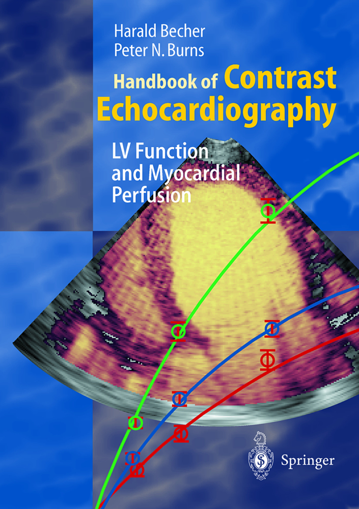
Handbook of Contrast Echocardiography
LV Function and Myocardial Perfusion. With a forew. by Sanjiv Kaul
Handbook of Contrast Echocardiography
LV Function and Myocardial Perfusion. With a forew. by Sanjiv Kaul
Although the technology required for the successful application of contrast echo cardiography has evolved rapidly over the past few years, the technique has not yet gained widespread clinical acceptance. One important reason for the lack of clinical acceptance is the relative complexity of the technique, particularly in respect to myocardial perfusion imaging. The interaction between micro bubbles and ultrasound is an entire field by itsel Pds. as is the coronary microvasculature. It is in this regard that practicing echocardiographers, cardiologists in training, radiologists, so no graphers, and students will find 'A Handbook of Contrast Echocardiography' particularly useful. Written by two leaders in the field who have presented illustrative cases not only from their own laboratories but also from others around the world, this volume is a lucid, concise, and practical guide for the day-to-day use of contrast echocardiography. Dr Peter Burns has been involved in almost all the technical advances in the imaging methods that have made it possible to detect opacification of the left ventricular cavity and myocardium from a venous injection of microbubbles. He has been responsible to a large degree for advancing our understanding of the interaction between micro bubbles and ultrasound, which he describes in clear and easy to understand terms in this book. Dr Harald Becher has been active in the clincal application of contrast echocardiography for several years and has gained considerable experience with many imaging techniques and microbubbles, which he describes in this volume in some detail.
1.2 Contrast agents for ultrasound
1.3 Mode of action
1.4 Safety considerations
1.5 New developments in contrast imaging
1.6 Summary
1.7 References
2 Assessment of Left Function Ventricular by Contrast Echo
2.1 Physiology and pathophysiology of LV function
2.2 Available methods - The role of contrast
2.3 Indications and selection of methods
2.4 How to perform an LV contrast study
2.5 Summary
2.6 References
3 Assessment of Myocardial Perfusion by Contrast Echo
3.1 Physiology and pathophysiology of myocardial perfusion
3.2 Currently available methods for myocardial perfusion imaging
3.3 Indications and selection of methods
3.4 Special considerations for myocardial contrast
3.5 Choice of agent and method of administration
3.6 Instrument settings
3.7 Image acquisition
3.8 Stress testing during myocardial contrast echo
3.9 Reading myocardial contrast echocardiograms
3.10 Clinical profiles/interpretation of myocardial contrast echo
3.11 Pitfalls and troubleshooting
3.12 Coronary flow reserve and myocardial contrast echo
3.13 Available methods - need for contrast enhancement
3.14 Coronary flow reserve: indications and selection of methods
3.15 How to perform a CFR study
3.16 Image acquisition and interpretation
3.17 Pitfalls and troubleshooting
3.18 Summary
3.19 References
4 Methods for quantitative Analysis
4.1 Basic tools for image quantification
4.2 Advanced image processing: cases & examples
4.3 Summary
4.4 References.
1 Contrast for agents echocardiography: Principles and Instrumentation
1.1 The need for contrast agents in echocardiography1.2 Contrast agents for ultrasound
1.3 Mode of action
1.4 Safety considerations
1.5 New developments in contrast imaging
1.6 Summary
1.7 References
2 Assessment of Left Function Ventricular by Contrast Echo
2.1 Physiology and pathophysiology of LV function
2.2 Available methods - The role of contrast
2.3 Indications and selection of methods
2.4 How to perform an LV contrast study
2.5 Summary
2.6 References
3 Assessment of Myocardial Perfusion by Contrast Echo
3.1 Physiology and pathophysiology of myocardial perfusion
3.2 Currently available methods for myocardial perfusion imaging
3.3 Indications and selection of methods
3.4 Special considerations for myocardial contrast
3.5 Choice of agent and method of administration
3.6 Instrument settings
3.7 Image acquisition
3.8 Stress testing during myocardial contrast echo
3.9 Reading myocardial contrast echocardiograms
3.10 Clinical profiles/interpretation of myocardial contrast echo
3.11 Pitfalls and troubleshooting
3.12 Coronary flow reserve and myocardial contrast echo
3.13 Available methods - need for contrast enhancement
3.14 Coronary flow reserve: indications and selection of methods
3.15 How to perform a CFR study
3.16 Image acquisition and interpretation
3.17 Pitfalls and troubleshooting
3.18 Summary
3.19 References
4 Methods for quantitative Analysis
4.1 Basic tools for image quantification
4.2 Advanced image processing: cases & examples
4.3 Summary
4.4 References.
Becher, Harald
Burns, Peter N.
Kaul, S.
| ISBN | 978-3-540-67083-4 |
|---|---|
| Artikelnummer | 9783540670834 |
| Medientyp | Buch |
| Copyrightjahr | 2000 |
| Verlag | Springer, Berlin |
| Umfang | XVI, 184 Seiten |
| Abbildungen | XVI, 184 p. 81 illus., 62 illus. in color. |
| Sprache | Englisch |

