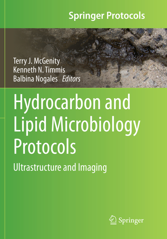
Hydrocarbon and Lipid Microbiology Protocols
Ultrastructure and Imaging
Hydrocarbon and Lipid Microbiology Protocols
Ultrastructure and Imaging
This Volume presents key microscopy and imaging methods for revealing the structure and ultrastructure of environmental and experimental samples, of microbial communities and cultures, and of individual cells. Method adaptations that specifically address problems concerning the hydrophobic components of samples are highlighted and discussed. The methods described range from electron microscopy and light and fluorescence microscopy, to confocal laser-scanning microscopy, and include experimental set-ups for the analysis of interfacial processes like microbial growth and activities at hydrocarbon:water interfaces, biofilms and microbe:mineral interfaces. Three forms of fluorescence in situ hybridization - CARD-FISH, MAR-FISH and Two-pass TSA-FISH - are described for the ecophysiological analysis of functionally active microbes in samples. The methods presented will enable readers to obtain an ultrastructural picture of, and identify the key functional microbes in, samples under investigation. This in turn will constitute a key framework for the interpretation of information from other experimental approaches, such as physicochemical analyses and genomic investigations.
Hydrocarbon and Lipid Microbiology ProtocolsThere are tens of thousands of structurally different hydrocarbons, hydrocarbon derivatives and lipids, and a wide array of these molecules are required for cells to function. The global hydrocarbon cycle, which is largely driven by microorganisms, has a major impact on our environment and climate. Microbes are responsible for cleaning up the environmental pollution caused by the exploitation of hydrocarbon reservoirs and will also be pivotal in reducing our reliance on fossil fuels by providing biofuels, plastics and industrial chemicals. Gaining an understanding of the relevant functions of the wide range of microbes that produce, consume and modify hydrocarbons and related compounds will be key to respondingto these challenges. This comprehensive collection of current and emerging protocols will facilitate acquisition of this understanding and exploitation of useful activities of such microbes.
Protocol for laser scanning microscopy of microorganisms on hydrocarbons
Fluorescence microscopy for microbiology
Imaging bacterial cells and biofilms adhering to hydrophobic organic compounds-water interfaces
Bacteria-mineral colloid interactions in biofilms - an ultrastructural and microanalytical approach
Identification of microorganisms in hydrocarbon contaminated aquifer samples by fluorescence in-situ hybridization (CARD-FISH)
Studies of the ecophysiology of single cells in microbial communities by (quantitative) microautoradiography and fluorescence in situ hybridization (MAR-FISH)
Protocol for in situ detection of functional genes of microorganisms by Two-pass TSA-FISH
Three-dimensional visualization and quantification of lipids in microalgae using confocal laser scanning microscopy
A Correlative Light-Electron Microscopy (CLEM) protocol for the identification of bacteria in animal tissue, exemplified by methanotrophic symbionts of deep-sea mussels.
Hydrocarbon and Lipid Microbiology ProtocolsThere are tens of thousands of structurally different hydrocarbons, hydrocarbon derivatives and lipids, and a wide array of these molecules are required for cells to function. The global hydrocarbon cycle, which is largely driven by microorganisms, has a major impact on our environment and climate. Microbes are responsible for cleaning up the environmental pollution caused by the exploitation of hydrocarbon reservoirs and will also be pivotal in reducing our reliance on fossil fuels by providing biofuels, plastics and industrial chemicals. Gaining an understanding of the relevant functions of the wide range of microbes that produce, consume and modify hydrocarbons and related compounds will be key to respondingto these challenges. This comprehensive collection of current and emerging protocols will facilitate acquisition of this understanding and exploitation of useful activities of such microbes.
Introduction
Electron microscopy protocols for the study of hydrocarbon-producing and -decomposing microbes: classical and advanced methodsProtocol for laser scanning microscopy of microorganisms on hydrocarbons
Fluorescence microscopy for microbiology
Imaging bacterial cells and biofilms adhering to hydrophobic organic compounds-water interfaces
Bacteria-mineral colloid interactions in biofilms - an ultrastructural and microanalytical approach
Identification of microorganisms in hydrocarbon contaminated aquifer samples by fluorescence in-situ hybridization (CARD-FISH)
Studies of the ecophysiology of single cells in microbial communities by (quantitative) microautoradiography and fluorescence in situ hybridization (MAR-FISH)
Protocol for in situ detection of functional genes of microorganisms by Two-pass TSA-FISH
Three-dimensional visualization and quantification of lipids in microalgae using confocal laser scanning microscopy
A Correlative Light-Electron Microscopy (CLEM) protocol for the identification of bacteria in animal tissue, exemplified by methanotrophic symbionts of deep-sea mussels.
McGenity, Terry J.
Timmis, Kenneth N.
Nogales, Balbina
| ISBN | 978-3-662-56983-2 |
|---|---|
| Artikelnummer | 9783662569832 |
| Medientyp | Buch |
| Auflage | Softcover reprint of the original 1st ed. 2016 |
| Copyrightjahr | 2018 |
| Verlag | Springer, Berlin |
| Umfang | X, 174 Seiten |
| Abbildungen | X, 174 p. |
| Sprache | Englisch |

