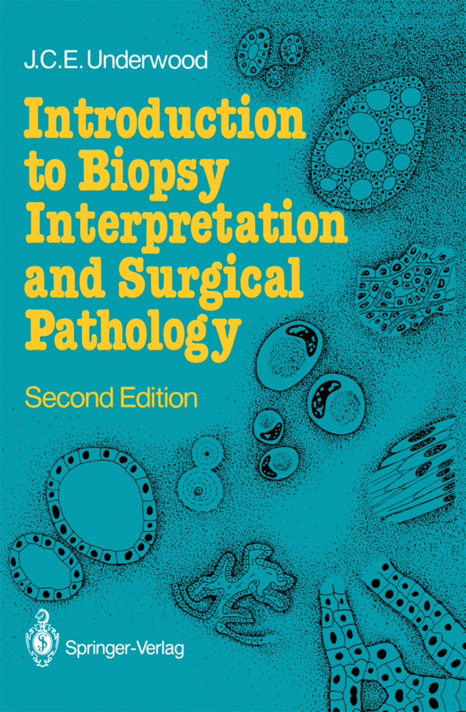
Introduction to Biopsy Interpretation and Surgical Pathology
Introduction to Biopsy Interpretation and Surgical Pathology
This is the second edition of the comprehensive and succinct account of basic principles and techniques of diagnostic histopathology, aimed at trainees in this discipline. Following a general introduction, the book gives a description of basic histopathological methods, such as tissue sampling and histological stains. Special techniques, including frozen sections, cytopathology, immunohistology, electron microscopy, and quantitive methods, are described in separate chapters. The methods of interpreting histological images and common causes of diagnostic difficulty are extensively illustrated. This new edition has been thoroughly revised and contains four new chapters, including quality control in histopathology and the autopsy. It is directly relevant to the activities of histopathologists and surgical pathologists, and will also be of value to physicians and surgeons in various specialties who use biopsies in clinical diagnosis.
1 Diagnostic Histopathology
Origins of HistopathologyThe Objectives of Histopathology
Medicolegal Aspects
The Histopathological Diagnosis
Adverse Effects of Biopsy
Prospects for Histopathology
2 Macroscopy, Microscopy and Sampling
Sampling Error
Sampling Biopsies and Surgical Resections
Specific Types of Biopsy
Specimen Radiology
Section Thickness
Imprints and Smears
Information and Magnification
3 The Use of Stains
Principles of Staining
Indications for Special Stains
Identification of Specific Substances and Cells
Autoradiography
4 Immunohistology
Techniques for Immunohistology
Primary Antibodies
Bridging Reagents
Tracer Substances
Fixation
Use of Enzymes to Unmask Antigens
Endogenous Peroxidase
Controls
Common Applications
Problems of Interpretation
Immuno-electron Microscopy
5 Interpretation of Histological Appearances
Artifact of Sections
Basic Microscopy
Artifacts in Sections
Methods of Interpretation
Clinicopathological Integration
Morphology of Disease Processes
6 Cytology
Relative Merits of Cytology and Histology
Cytological Preparations
Stains for Cytology
Special Investigations
Interpretation
Applications
Living Cells
Reporting Cytology
7 Borderline Lesions, Pseudomalignancy and Mimicry
Borderline Lesions
Mimicry of Histological Features
Pseudomalignancy and Related Diagnostic Problems
Conclusions
8 Rapid Frozen Section Diagnosis
Clinical Indications
Methods
Interpretation
Common Applications
Reliability
9 Diagnostic Electron Microscopy
Principles of Transmission Electron Microscopy
Diagnostic Value of Electron Microscopy
Scanning Electron Microscopy
X-ray Microanalysis
10 Quantitative Methods
Stereological Principles
Stereological Methods
Automatic and Semi-automatic Image Analysis
Microspectrophotometry
Flow Cytometry
Practical Applications
11 Reporting and Classification of Biopsy Diagnoses
The Biopsy Report
Disease Classification
Data Storage and Retrieval
Clinical Assimilation
12 Quality Assessment and Control
Sources of Diagnostic Error
The "Correct" Diagnosis
Evaluation of Diagnostic Methods
Evaluation of Performance
Illustrative Examples
Self Assessment
Assessment of Errors of Procedure and Communication
Conclusions
13 The Autopsy
History of the Autopsy
The Autopsy and Clinical Audit
Role in Medical Education
Hazards in the Autopsy Room
Consent
General Techniques
Special Methods
Post-mortem Histology.
Contents: Diagnostic Histopathology
Macroscopy, Microscopy and SamplingThe Use of Stains
Immunohistology
Interpretation of Histological Appearances
Cytology
Borderline Lesions, Pseudomalignancy and Mimicry
Rapid Frozen Section Diagnosis
Diagnostic Electron Microscopy
Quantitative Methods
Reporting and Classification of Biopsy Diagnoses
Quality Assessment and Control
The Autopsy
Subject Index.
Underwood, James C. E.
| ISBN | 978-3-540-17495-0 |
|---|---|
| Artikelnummer | 9783540174950 |
| Medientyp | Buch |
| Auflage | 2. Aufl. |
| Copyrightjahr | 1987 |
| Verlag | Springer, Berlin |
| Umfang | XIV, 218 Seiten |
| Abbildungen | XIV, 218 p. 63 illus. |
| Sprache | Englisch |

