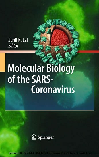
SARS was the ?rst new plague of the twenty-?rst century. Within months, it spread worldwide from its 'birthplace' in Guangdong Province, China, affecting over 8,000 people in 25 countries and territories across ?ve continents. SARS exposed the vulnerability of our modern globalised world to the spread of a new emerging infection. SARS (or a similar new emerging disease) could neither have spread so rapidly nor had such a great global impact even 50 years ago, and arguably, it was itself a product of our global inter-connectedness. Increasing af?uence and a demand for wild-game as exotic food led to the development of large trade of live animal and game animal markets where many species of wild and domestic animals were co-housed, providing the ideal opportunities for inter-species tra- mission of viruses and other microbes. Once such a virus jumped species and attacked humans, the increased human mobility allowed the virus the opportunity for rapid spread. An infected patient from Guangdong who stayed for one day at a hotel in Hong Kong led to the transmission of the disease to 16 other guests who travelled on to seed outbreaks of the disease in Toronto, Singapore, and Vietnam, as well as within Hong Kong itself. The virus exploited the practices used in modern intensive care of patients with severe respiratory disease and the weakness in infection control practices within our health care systems to cause outbreaks within hospitals, further amplifying the spread of the disease. Health-care itself has become a two-edged sword.
1;Foreword;5 2;Preface;7 3;Contents;10 4;Part I: Viral Entry;13 4.1;Chapter 1: Cellular Entry of the SARS Coronavirus: Implications for Transmission, Pathogenicity and Antiviral Strategies;14 4.1.1;Introduction;14 4.1.2;The Spike Protein: Key to the Host Cell;15 4.1.3;The Attachment Factors DC-SIGN and DC-SIGNR: Enhancers or Inhibitors of SARS-CoV Infection?;17 4.1.4;The Two Faces of ACE2: SARS-CoV Receptor and Protector Against Lung Damage;18 4.1.4.1;The Structure of the Interface Between SARS-S and ACE2;19 4.1.4.2;Sequence Variations at the SARS-S/ACE2 Interface Might Impact Viral Transmission and Pathogenicity;20 4.1.4.3;The Human Coronavirus NL63 Uses ACE2 for Cellular Entry;21 4.1.4.4;SARS Versus NL63: A Correlation Between ACE2 Downregulation and Viral Pathogenicity?;21 4.1.5;Cleavage by Endosomal Cathepsin Proteases Activates SARS-S;23 4.1.6;Membrane Fusion is Driven by Conserved Elements Located in the S2 Subunit of the SARS Spike Protein;24 4.1.7;Conclusions;26 4.1.8;References;26 4.2;Chapter 2: The Cell Biology of the SARS Coronavirus Receptor, Angiotensin-Converting Enzyme 2;34 4.2.1;Introduction;34 4.2.2;Clues from Homologous Proteins;35 4.2.3;Regulation of ACE2 Expression on the Cell Surface;37 4.2.3.1;Proteolytic Cleavage Secretion;37 4.2.3.2;The Role of Membrane Microdomains;38 4.2.4;Conclusions and Future Perspectives;39 4.2.5;References;39 4.3;Chapter 3: Structural Molecular Insights into SARS Coronavirus Cellular Attachment, Entry and Morphogenesis;42 4.3.1;Structure of SARS Coronavirus (SARS-CoV);43 4.3.2;Structure of the Coronavirus Spike;45 4.3.3;Viral Membrane Fusion in SARS-CoV;47 4.3.4;Cellular Attachment and Entry of SARS-CoV;47 4.3.5;References;52 5;Part II: Structures Involved in Viral Replication and Gene Expression;55 5.1;Chapter 4: RNA Higher-Order Structures Within the Coronavirus 5 and 3 Untranslated Regions and Their Roles in Viral Replica;56 5.1.1;Introduction;56 5.1.2;cis-Acting RNA Elements in Coronavirus Replication;57 5.1.2.1;The Transcription Regulatory Sequence;58 5.1.2.2;The 5 cis-Acting RNA Elements;59 5.1.2.3;The 3 cis-Acting RNA Elements;62 5.1.2.4;Proteins Binding to the 5 and 3 cis-Acting Elements;64 5.1.3;Future Directions;66 5.1.4;References;66 5.2;Chapter 5: Programmed -1 Ribosomal Frameshifting in SARS Coronavirus;71 5.2.1;Introduction;71 5.2.2;Programmed -1 Ribosomal Frameshifting;72 5.2.3;Programmed Frameshifting Rates and Virus Propagation;73 5.2.4;Different Models, Different Assay Systems, Different Results;74 5.2.5;The Biology of -1 PRF in SARS-CoV is Different;75 5.2.6;A Unique Feature of the SARS-CoV Frameshift Signal: A Three-Stemmed mRNA Pseudoknot;76 5.2.7;A Second PRF Signal in SARS-CoV?;77 5.2.8;Summary and Perspectives;78 5.2.9;References;78 6;Part III: Viral Proteins;81 6.1;Chapter 6: Expression and Functions of SARS Coronavirus Replicative Proteins;82 6.1.1;Introduction;82 6.1.2;Organization and Expression of the Coronavirus Replicase Gene;85 6.1.3;Functions and Activities of Replicase Gene-Encoded Nonstructural Proteins;86 6.1.3.1;ORF1a-Encoded Nonstructural Proteins 1-11;86 6.1.3.2;ORF1b-Encoded Nonstructural Proteins 12-16;93 6.1.4;Future Directions;97 6.1.5;References;99 6.2;Chapter 7: SARS Coronavirus Replicative Enzymes: Structures and Mechanisms;106 6.2.1;The ADP-Ribose-1-Phosphatase Domain;107 6.2.2;The nsp7-nsp8-nsp9-nsp10 Cistron;109 6.2.2.1;The nsp7 Protein;109 6.2.2.2;The nsp8 Protein;110 6.2.2.3;The nsp9 Protein;113 6.2.2.4;The nsp10 Protein;115 6.2.3;The Endoribonuclease, nsp15;116 6.2.4;Conclusion;118 6.2.5;References;119 6.3;Chapter 8: Quaternary Structure of the SARS Coronavirus Main Protease;122 6.3.1;Introduction;123 6.3.2;Molecular Biology of the SARS-CoV Polyproteins;123 6.3.3;Structure of the SARS-CoV Main Protease;124 6.3.3.1;Three-Dimensional Structure of the SARS-CoV Main Protease;124 6.3.3.2;Quaternary StructureMproQuaternary structure of the SARS-CoV Main Protease;126 6.3.4;Enzyme Activity-AssayMproactivity-assay for the SARS-CoV Main P
Lal, Sunil K.
| ISBN | 9783642036835 |
|---|---|
| Artikelnummer | 9783642036835 |
| Medientyp | E-Book - PDF |
| Auflage | 2. Aufl. |
| Copyrightjahr | 2010 |
| Verlag | Springer-Verlag |
| Umfang | 328 Seiten |
| Sprache | Englisch |
| Kopierschutz | Digitales Wasserzeichen |

