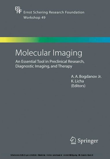
Molecular Imaging
An Essential Tool in Preclinical Research, Diagnostic Imaging, and Therapy
The continuous progress in the understanding of molecular processes of disease formation and progression attributes an increasing importance to biomedical molecular imaging methods. The purpose of this workshop was to discuss and overview multiple applications and emerging technologies in the area of diagnostic imaging including its fundamental capabilities in preclinical research, the opportunities for medical care, and the options involving therapeutic concepts. The book provides the reader with state-of-the-art information on the different aspects of diagnostic imaging, illuminating new developments in molecular biology, imaging agents and molecular probe design, and therapeutic techniques.
1;Preface;5 2;Contents;7 3;List of Editors and Contributors;9 4;1 Oligonucleotides as Radiopharmaceuticals;13 4.1;1.1 Imaging of Oligonucleotides;17 4.2;1.2 Imaging In Vivo Pharmacokinetics and Biodistribution of Oligonucleotides;20 4.3;1.3 Comparative Pharmaco-Imaging of Oligonucleotides;21 4.4;1.4 Evaluation of Delivery Vectors for Oligonucleotides;23 4.5;1.5 Imaging with Oligonucleotides;25 4.6;1.6 Antisense Oligonucleotides for Gene-Related Disease Therapy;26 4.7;1.7 Improving Antisense for In Vivo Applications;27 4.8;1.8 In Vivo Imaging Studies with Antisense Oligonucleotides;28 4.9;1.9 Aptamer Oligonucleotides;30 4.10;1.10 Applications and Therapeutic Aptamers;33 4.11;1.11 Aptamers as Contrast Agents for Diagnosis;34 4.12;1.12 Future Developments Needed for In Vivo Imaging with Oligonucleotides: Mastering the Nonspecific Interactions and Signal-to-Noise Ratio;35 4.13;References;38 5;2 ImagingProtein-Protein Interactions in Whole Cells and Living Animals;47 5.1;2.1 Two-Hybrid Systems;48 5.2;2.2 Protein-Fragment Complementation;49 5.3;2.3 Optimized Luciferase Fragment Complementation;50 5.4;2.4 Conclusions;51 5.5;References;52 6;3 Radiolabeled Peptides in Nuclear Oncology: Influence of Peptide Structure and Labeling Strategy on Pharmacology;54 6.1;3.1 Introduction;54 6.2;3.2 Design of Peptide-Based Radiopharmaceuticals;58 6.3;3.3 Preclinical Characterization of Somatostatin-Based Radiopeptides;69 6.4;3.4 Patient Studies;77 6.5;3.5 Summary and Conclusions;80 6.6;References;80 7;4 Pretarieted Radioimmunotherapy;84 8;5 PET/CT:Combining Function and Morphology;96 8.1;5.1 General Considerations in PET and PET/CT;96 8.2;5.2 PET/CT in Oncology;98 8.3;5.3 PET/CT and Radiation Treatment Planning;104 8.4;5.4 PET/CT in Cardiology;104 8.5;5.5 Conclusions;106 8.6;References;107 9;6 High Relaxivity Contrast Agents for MRI and Molecular Imaging;110 9.1;6.1 Introduction;110 9.2;6.2 Determinants of Relaxivity;114 9.3;6.3 How to Improve Relaxivity?;116 9.4;6.4 Targeting Cells with Gd(III) Chelates;122 9.5;6.5 Concluding Remarks;129 9.6;References;130 10;7 Luminescent Lanthanide Complexes as Sensors and Imaiini Proies;133 10.1;7.1 Introduction and Background;133 10.2;7.2 Mechanistic Approach to Modulation of Luminescence;137 10.3;7.3 Tailoring the Lanthanide Complex for Use "In Cellulo";144 10.4;7.4 Conclusions and Future Perspectives;152 10.5;References;153 11;8 Mainetic Resonance Signal Amplification Probes;157 11.1;8.1 Receptor-Mediated Internalization;158 11.2;8.2 Enzyme-Mediated MR Signal Amplification;162 11.3;References;166 12;9 Imaging of Proteases for Tumor Detection and Differentiation;168 12.1;9.1 Introduction;168 12.2;9.2 Light for Molecular Imaging;169 12.3;9.3 Optical Contrast Agents;170 12.4;9.4 Nononcological Imaging;174 12.5;9.5 Potential Clinical Applications;175 12.6;References;177 13;10 Molecular Imaging with Targeted Ultrasound Contrast Microiuiiles;180 13.1;10.1 Introduction;180 13.2;10.2 Microbubble-Based Ultrasound Contrast Agents: General Design and Preparation;182 13.3;10.3 Targeting Ligand Attachment to Microbubbles;185 13.4;10.4 In Vitro Targeting: Binding Studies in Model Systems;189 13.5;10.5 In Vivo Microbubble Targeting: Biodistribution, Nonspecific and Targeted Accumulation and Ultrasound Imaging;192 13.6;10.6 Conclusions;197 13.7;References;198 14;11 Noninvasive Real-Time In Vivo Bioluminescent Imaging of Gene Expression and of Tumor Progression and Metastasis;201 14.1;11.1 General Introduction;202 14.2;11.2 Principles of Bioluminescent Imaging;202 14.3;11.3 Transgenic Luc-Reporter Mice to Study Specific Gene Expression;204 14.4;11.4 Tumor Progression and Bone/Bone Marrow Metastasis of Breast and Prostate Cancer;213 14.5;11.5 Use of Bioluminescent Imaging in Animal Models of Skeletal Metastasis;220 14.6;11.6 Conclusions and Future Perspectives;228 14.7;References;228 15;12 Tarieted Optical Imaging and Photodynamic Therapy;236 15.1;12.1 Introduction;236 15.2;12.2 Photosensitizer Localization in Tumors;238 15.
| ISBN | 9783540268093 |
|---|---|
| Artikelnummer | 9783540268093 |
| Medientyp | E-Book - PDF |
| Copyrightjahr | 2007 |
| Verlag | Springer-Verlag |
| Umfang | 258 Seiten |
| Sprache | Englisch |
| Kopierschutz | Digitales Wasserzeichen |

