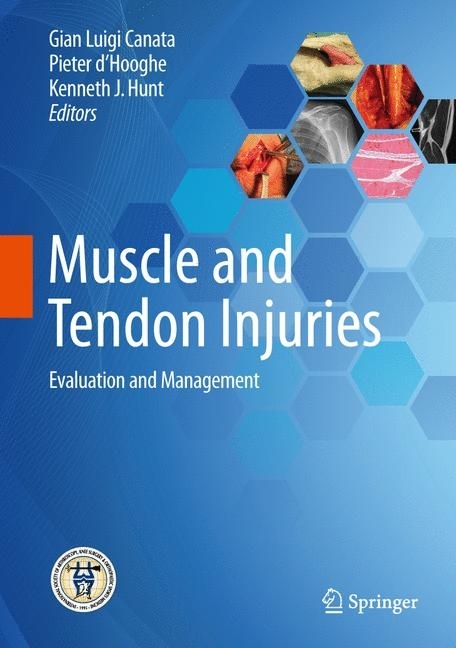
Muscle and Tendon Injuries
Evaluation and Management
This book explores in a comprehensive manner the causes and symptoms of muscle and tendon pathologies, the available diagnostic procedures, and current treatment approaches. Specific aspects of the anatomy, biomechanics, and function of muscles and tendons are analyzed, and detailed guidance is provided on the most innovative methods - both conservative and surgical - for ensuring that the athlete can make a safe and quick return to sporting activity. Optimal care of tendon and muscle injuries in sportspeople requires effective cooperation of sports scientists and medical practitioners to identify the best ways of preserving muscle and tendon structures and to develop new strategies for their rehabilitation and regeneration. Muscle and Tendon Injuries is an excellent multidisciplinary reference written by the leading experts in the field and published in collaboration with ISAKOS. It will appeal to all specialists in sports medicine and sports traumatology who are seeking a state of the art update on the management of muscle and tendon disorders.
Gian Luigi Canata, MD, has been Director of the Center of Sports Traumatology and Arthroscopic Surgery at Koelliker Hospital, Turin, Italy since 1995. He has been a member of the Medical Commission of the Italian Track and Field Federation (FIDAL) and Director of Medical Services and a member of the Board of Directors of the Centro Universitario Sportivo of Torino (CUS Torino) for more than 35 years. His previous academic positions include Professor of Sports Medicine at SUISM (Scuola Universitaria in Scienze Motorie) (2001-2012) and Professor of Kinesiology at ISEF (Istituto Superiore di Educazione Fisica) (1989-2001). Dr. Canata has been a member of ISAKOS since 1995 and has served on various ISAKOS committees. He is also President of the SIGASCOT Sports Committee and Vice-President of EFOST. He has authored almost 120 articles, many of them in peer-reviewed journals and is co-editor, with C. Niek Van Dijk, of the 2015 Springer book Cartilage Lesions of the Ankle.
Pieter d'Hooghe is Associate Chief of Surgery (Research) at the Department of Orthopaedic Surgery, Aspetar Hospital, Qatar. As an orthopaedic surgeon, he specializes in arthroscopic surgery of the ankle and foot, knee, and hip. He holds a Master in Sports Medicine and Tropical Medicine and has an MBA on Leadership and Sports Management. He was previously Director of the FIFA F-MARC Flemish Surgical Center of Excellence, where he treated many national and international athletes. Prior to working in Aspetar, Dr. d'Hooghe spent 17 years as team physician/surgeon of FC Club Bruges in Belgium (Champions League, Europa League, 7 national titles, 4 national cups, 3 national supercups). As Chairman of the ISAKOS Leg, Ankle, and Foot Committee he heads an international group of experts in the field, and as FIFA F-MARC Board Member and Instructor he is also involved in research, injury prevention, delivering lectures worldwide, publishing peer-reviewed articles, books and book chapters and assisting in PCMA projects.
Kenneth J. Hunt, MD, is Associate Professor and Chief, Foot and Ankle Surgery in the Department of Orthopedics, University of Colorado School of Medicine, Aurora, CO, USA. He was previously Assistant Professor in the Department of Orthopaedic Surgery at Stanford University and Director of the Stanford/Palo Alto Foot & Ankle Fellowship. Dr. Hunt is Deputy Chair of the ISAKOS Leg, Ankle, and Foot Committee. He also chairs the Orthopaedic Foot and Ankle Research Network (OFAR) and is a member of the Board of Directors of the Northern California Orthopaedic Society. Dr. Hunt is a Diplomate of the American Board of Orthopaedic Surgeons. He is an Associate Editor of Physical Medicine & Rehabilitation and a reviewer for various leading journals, including the American Journal of Sports Medicine and the Journal of Bone and Joint Surgery.
1;Foreword;5 2;Preface;6 3;Acknowledgements;7 4;Contents;3 5;1: Functional Morphology of Muscles and Tendons;11 5.1;1.1 Muscle;11 5.1.1;1.1.1 Anatomy of Skeletal Muscle Tissue;12 5.1.2;1.1.2 Classification of Skeletal Muscles;14 5.1.3;1.1.3 Number of Points of Origin;15 5.1.4;1.1.4 Mode of Action;15 5.1.5;1.1.5 Classification of Skeletal Muscle Fibres;16 5.1.6;1.1.6 Motor Units and Their Recruitment;18 5.1.7;1.1.7 Vascularisation and Innervation;18 5.1.8;1.1.8 Muscle Plasticity;19 5.2;1.2 Tendons;19 5.2.1;1.2.1 Tendon Structure;19 5.2.2;1.2.2 Biochemical Composition of the Tendon;20 5.2.2.1;1.2.2.1 Collagen;21 5.2.2.2;1.2.2.2 Elastin;21 5.2.2.3;1.2.2.3 Proteoglycans;21 5.2.3;1.2.3 Tendon Cells;22 5.2.4;1.2.4 Tendon Crimps;22 5.2.5;1.2.5 Vascularisation;23 5.2.6;1.2.6 Innervation;23 5.3;References;23 6;2: Tendon Biomechanics;25 6.1;2.1 Introduction;25 6.2;2.2 Composition and Structure;25 6.3;2.3 Mechanics;26 6.4;2.4 Mechanical Testing Methods;29 6.5;2.5 Experimental Factors Influencing Tendon Mechanics;30 6.6;2.6 Biological Factors Influencing Tendon Mechanics;31 6.7;References;32 7;3: Pathophysiology of Tendinopathy;33 7.1;3.1 Structure and Histology;33 7.2;3.2 Tenocytes;34 7.3;3.3 Extracellular Matrix;34 7.3.1;3.3.1 Ground Substance;35 7.4;3.4 Blood and Nerve Supply;35 7.5;3.5 Structural Regions in Tendons;36 7.6;3.6 Response to Load;36 7.7;3.7 Function and Mechanics of Tendons;37 7.8;3.8 Summary;37 7.9;3.9 Structural Changes in Tendon Pathology;37 7.10;3.10 Pathoaetiology;38 7.10.1;3.10.1 Continuum Model of Tendinopathy;38 7.11;3.11 Pain in Tendinopathy;39 7.12;3.12 Regional Changes in Tendinopathy;40 7.13;3.13 Epidemiology of Tendinopathy;40 7.14;3.14 Intrinsic Risk Factors;41 7.14.1;3.14.1 Genetics;42 7.14.2;3.14.2 Sex;42 7.14.3;3.14.3 Age;42 7.14.4;3.14.4 Medications;43 7.14.5;3.14.5 General Health;43 7.14.6;3.14.6 Flexibility and Joint Stiffness;44 7.14.7;3.14.7 Strength;44 7.15;3.15 Extrinsic Risk Factors;45 7.15.1;3.15.1 Load;45 7.16;References;45 8;4: Tendon Healing;55 8.1;4.1 Introduction;55 8.2;4.2 Tendon Homeostasis;55 8.2.1;4.2.1 Mechanism of Tendon Injury;56 8.2.1.1;4.2.1.1 Stages of Tendon Healing;56 8.2.1.2;4.2.1.2 Growth Factors;57 8.2.1.3;4.2.1.3 Tendon Healing of Intrasynovial and Extrasynovial Tendon;57 8.3;4.3 Rehabilitation;58 8.4;References;59 9;5: Muscle-Tendon Junction Injury;61 9.1;5.1 Introduction;61 9.2;5.2 Structure;61 9.3;5.3 Function;63 9.4;5.4 Injury;64 9.5;5.5 Recovery;66 9.6;5.6 Clinical Implications;67 9.7;References;69 10;6: MRI of Muscle and Tendon Pathology;71 10.1;6.1 Introduction;71 10.2;6.2 Standard MRI;72 10.2.1;6.2.1 Contrast-Enhanced MRI;72 10.2.2;6.2.2 Magnetic Resonance Angiography;72 10.2.3;6.2.3 Fusion Technology;73 10.2.4;6.2.4 Diffusion MRI;74 10.2.5;6.2.5 MR Elastography;74 10.2.6;6.2.6 Upright MRI;74 10.2.7;6.2.7 Dynamic MRI;75 10.3;6.3 Muscles;75 10.3.1;6.3.1 Trauma-Related Conditions;75 10.3.1.1;6.3.1.1 Minor Traumas;75 10.3.1.2;6.3.1.2 Major Traumas;76 10.3.2;6.3.2 Myofascial Disinsertion;77 10.3.3;6.3.3 Myotendinous Avulsion;77 10.3.4;6.3.4 Lesions Due to Compression Injury;77 10.3.5;6.3.5 Post-traumatic Degenerative Conditions;78 10.3.5.1;6.3.5.1 Post-traumatic Fibrosis;78 10.3.5.2;6.3.5.2 Metaplastic Ossification;79 10.3.5.3;6.3.5.3 Rhabdomyolysis;79 10.3.6;6.3.6 Compartmental Syndromes;79 10.3.6.1;6.3.6.1 Muscle Hernias;80 10.3.6.2;6.3.6.2 Neuromuscular Conditions;80 10.3.7;6.3.7 Neoplastic Disease;80 10.4;6.4 Tendons;80 10.4.1;6.4.1 Peritendinitis;81 10.4.2;6.4.2 Degenerative Tendinopathy;82 10.4.3;6.4.3 Partial Tears;82 10.4.4;6.4.4 Complete Tears;82 10.4.5;6.4.5 Tenosynovitis;82 10.4.6;6.4.6 Tenorrhaphy;83 10.5;Suggested Reading;83 11;7: Ultrasound of Muscle and Tendon Pathology;84 11.1;7.1 Introduction;84 11.2;7.2 B-Mode Ultrasound;84 11.3;7.3 Ultrasound Fusion;85 11.4;7.4 Elastography;85 11.5;7.5 Muscles;86 11.5.1;7.5.1 Trauma-Related Conditions;86 11.5.1.1;7.5.1.1 Minor Traumas;86 11.5.1.2;7.5.1.2 Major Traumas;86 11.5.2;7.5.2 Complete Ruptures;88 11.5.3;7.5.3 My
Canata, Gian Luigi
Hunt, Kenneth J.
| ISBN | 9783662541845 |
|---|---|
| Artikelnummer | 9783662541845 |
| Medientyp | E-Book - PDF |
| Copyrightjahr | 2017 |
| Verlag | Springer-Verlag |
| Umfang | 440 Seiten |
| Sprache | Englisch |
| Kopierschutz | Digitales Wasserzeichen |

