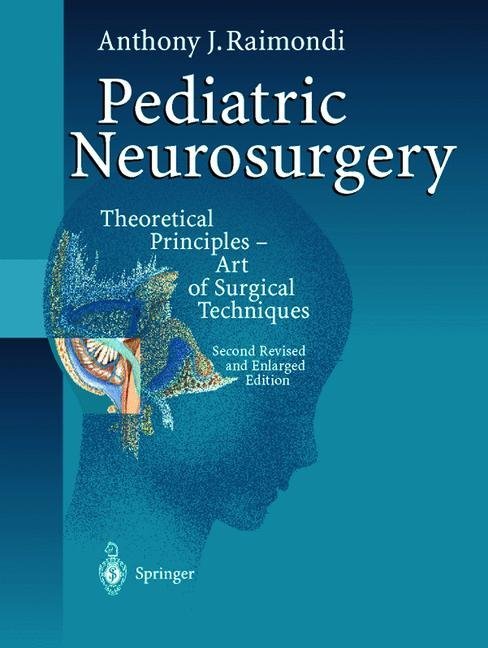
Pediatric Neurosurgery
Theoretical Principles - Art of Surgical Techniques
Pediatric Neurosurgery
Theoretical Principles - Art of Surgical Techniques
Both a theoretic text-book and a descriptive atlas, this standard reference in the field of pediatric neurosurgery presents basic clinical concepts and surgical techniques in a step-by-step fashion. The neuro-imaging essential both to clinical diagnosis and surgical planning are set into the text in a consequential manner, endeavoring to facilitate visual retention and spatial orientation. The illustrations present the visual perspectives of the surgeon, they are often integrated into imaging studies, and the sequence of their presentation is designed to permit time-frame comprehension. Great attention is given to the dynamics of decision making (offering alternatives) and where particular caution is the pass-word. The different perspectives and opinions of other leaders in the field are woven into the discussions, developing for the reader concepts upon which he may draw. The review of the world literature between 1980 and 1998 is complete, but the text is not laden with citations, quotes, supporting and opposing views. The author expresses these throughout the book as the synthesis of his readings, discussions, experiences, and reflections.
General Discussion
Specific Positions
Positioning of the Child Vis-à-vis the Surgeon's Line of Sight
Positions of Surgeon, Assistants, and Nurse Around the Patient
Accommodating Anesthesia
General Positions
Supine Position
Prone Position
Lounging Position
References
2 Incisions: Scalp, Muscle, Tissue, and Tumor Hemostasis
Specific Incisions for Surgical Approaches
Bifrontal Incision
Frontal Incision
Frontoparietal Skin Incision for Frontoparietal Bur Holes
Frontoparietal Incision for Posterofrontal or Anteroparietal Lesions
Parietal Incision
Parasagittal Incision
Temporal Incision
Occipital Incision
Suboccipital Incision
Combined Supra-and Infratentorial Incision
Hemispherical Incision
Laminotomy
Techniques for Scalp Hemostasis in Various Ages: Newborn, Infant, Toddler
Specific Types of Bleeding
Closure
Laminotomy
3 Bur Holes and Flaps
Bur Holes: Frontoparietal (So-Called "Diagnostic")
Flaps
Bifrontal Flap
Frontal Flap
Approaches to the Orbit
Parietal Flap
Parietotemporal Flap
Biparietal Craniotomy
Temporal Flaps
Occipital Flaps
Suboccipital Flaps
Supra-and Infratentorial Craniotomy
Hemispherical Craniotomy
Laminotomy
Bone Closure
References
4 Suturotomy for Various Flaps in the Newborn and Infant
5 Dural Flaps
General Comments
Dural Openings
Closure
References
6 Cerebral Retraction
Cistern Openings
Use of Gravity
Ambient Cistern Lesions
Use of Cotton Fluffies and Telfa
Self-Retaining Retractors
References
7 Cerebrotomy
Gyral Cerebrotomy
Sulcal Cerebrotomy
Small Vessels at the Depth of the Sulcus or Gyrus
Cerebrotomy Through White Matter
8 Cerebral Resection
Biopsy
Lobectomy
Hemispherectomy
9Epilepsy
Results
Definitions
Epidemiology
Classification of Epileptic Seizures
Patient Selection
Noninvasive Methodologies
History and Physical
EEG and Video- EEG
Diagnosis by Imaging Studies
Invasive Methodologies
Operative Procedures
References
10 Tumors
Surgical Approach and Removal
Bone Tumors
Age and Brain Tumors
Materials and Methods
Hemispherical Tumors
Ventricular Tumors
Vermian Tumors
Foramen Magnum Tumors
Cerebellar Hemisphere Tumors
Parasellar Tumors
Corpus Callosum and Septum Pellucidum Tumors
Spinal Tumors
Intradural-Extramedullary Tumors
Intramedullary Tumors
References
11 Vascular Disorders: Surgical Approaches and Operative Technique
Saccular Aneurysms
Vascular Malformations
Transcranial Venovenous Shunts
Transcranial Arteriovenous Fistulae
Dural Arteriovenous Fistulae
Superior Sagittal Sinus Thrombosis
Parenchymal Arteriovenous Malformations
Venous Angiomas
Cerebellar Hemisphere
Arteriovenous Malformations
Orbital Cavernoma
Brainstem Arteriovenous Malformations
Spontaneous Intraparenchymal Hemorrhage
Intraventricular Arteriovenous Malformations
Arteriovenous Fistulae Involving Galenic System and/or Perimesencephalic Leptomeninges
Heart Failure, Arteriovenous Shunting, Thrombophlebitis, and Hydrocephalus
Endovascular Occlusion or Open Surgery
References
12 Infections
Osteomyelitis
Epidural Empyema
Subdural Abscess and Subdural Empyema
Acute Meningitis with Hydrocephalus
Brain Abscess
Bur Hole and Cannula Drainage
Craniotomy and Resection of the Abscess
Pyocephalus
Ventriculitis
Subdural Effusions
Cerebritis and Cerebellitis
Wound Infections
References
13 Trauma
Injuries of the Scalp
Cerebral Contusion and Edema
Epidural Hematoma
Cerebrospinal Fluid Leaks
Meningocele Spuria
Child Abuse
Post -traumatic Cerebrovascular Injuries
False Aneurysms
Cranioplasty
Vertebral Fracture Dislocation
References
14 Congenital Anomalies
Congenital Anomalies Involving the Craniocerebrum and Craniocervical Junction
Hypertelorism
Arachnoidal Cysts
Craniofacial Encephalomeningoceles
Cranioschisis
Cranial Meningoencephalocele
Craniocerebral Disproportions: Chiari Malformations
Separation of Craniopagus Twins
Vertebrospinal Congenital Anomalies
The Dysraphic State
Nondysraphic Spinal Cord Anomalies and Congenital Tumors
References
15 Hydrocephalus
Definition and Classification
Surgical Treatment and Prognosis
Genesis of Parenchymal Destruction
Diagnosis
Treatment
Intellectual Development and Quality of Survival
Surgical Management
Techniques for Cannulation of the Ventricles
Shunts
References.
List of Contents
1 PositioningGeneral Discussion
Specific Positions
Positioning of the Child Vis-à-vis the Surgeon's Line of Sight
Positions of Surgeon, Assistants, and Nurse Around the Patient
Accommodating Anesthesia
General Positions
Supine Position
Prone Position
Lounging Position
References
2 Incisions: Scalp, Muscle, Tissue, and Tumor Hemostasis
Specific Incisions for Surgical Approaches
Bifrontal Incision
Frontal Incision
Frontoparietal Skin Incision for Frontoparietal Bur Holes
Frontoparietal Incision for Posterofrontal or Anteroparietal Lesions
Parietal Incision
Parasagittal Incision
Temporal Incision
Occipital Incision
Suboccipital Incision
Combined Supra-and Infratentorial Incision
Hemispherical Incision
Laminotomy
Techniques for Scalp Hemostasis in Various Ages: Newborn, Infant, Toddler
Specific Types of Bleeding
Closure
Laminotomy
3 Bur Holes and Flaps
Bur Holes: Frontoparietal (So-Called "Diagnostic")
Flaps
Bifrontal Flap
Frontal Flap
Approaches to the Orbit
Parietal Flap
Parietotemporal Flap
Biparietal Craniotomy
Temporal Flaps
Occipital Flaps
Suboccipital Flaps
Supra-and Infratentorial Craniotomy
Hemispherical Craniotomy
Laminotomy
Bone Closure
References
4 Suturotomy for Various Flaps in the Newborn and Infant
5 Dural Flaps
General Comments
Dural Openings
Closure
References
6 Cerebral Retraction
Cistern Openings
Use of Gravity
Ambient Cistern Lesions
Use of Cotton Fluffies and Telfa
Self-Retaining Retractors
References
7 Cerebrotomy
Gyral Cerebrotomy
Sulcal Cerebrotomy
Small Vessels at the Depth of the Sulcus or Gyrus
Cerebrotomy Through White Matter
8 Cerebral Resection
Biopsy
Lobectomy
Hemispherectomy
9Epilepsy
Results
Definitions
Epidemiology
Classification of Epileptic Seizures
Patient Selection
Noninvasive Methodologies
History and Physical
EEG and Video- EEG
Diagnosis by Imaging Studies
Invasive Methodologies
Operative Procedures
References
10 Tumors
Surgical Approach and Removal
Bone Tumors
Age and Brain Tumors
Materials and Methods
Hemispherical Tumors
Ventricular Tumors
Vermian Tumors
Foramen Magnum Tumors
Cerebellar Hemisphere Tumors
Parasellar Tumors
Corpus Callosum and Septum Pellucidum Tumors
Spinal Tumors
Intradural-Extramedullary Tumors
Intramedullary Tumors
References
11 Vascular Disorders: Surgical Approaches and Operative Technique
Saccular Aneurysms
Vascular Malformations
Transcranial Venovenous Shunts
Transcranial Arteriovenous Fistulae
Dural Arteriovenous Fistulae
Superior Sagittal Sinus Thrombosis
Parenchymal Arteriovenous Malformations
Venous Angiomas
Cerebellar Hemisphere
Arteriovenous Malformations
Orbital Cavernoma
Brainstem Arteriovenous Malformations
Spontaneous Intraparenchymal Hemorrhage
Intraventricular Arteriovenous Malformations
Arteriovenous Fistulae Involving Galenic System and/or Perimesencephalic Leptomeninges
Heart Failure, Arteriovenous Shunting, Thrombophlebitis, and Hydrocephalus
Endovascular Occlusion or Open Surgery
References
12 Infections
Osteomyelitis
Epidural Empyema
Subdural Abscess and Subdural Empyema
Acute Meningitis with Hydrocephalus
Brain Abscess
Bur Hole and Cannula Drainage
Craniotomy and Resection of the Abscess
Pyocephalus
Ventriculitis
Subdural Effusions
Cerebritis and Cerebellitis
Wound Infections
References
13 Trauma
Injuries of the Scalp
Cerebral Contusion and Edema
Epidural Hematoma
Cerebrospinal Fluid Leaks
Meningocele Spuria
Child Abuse
Post -traumatic Cerebrovascular Injuries
False Aneurysms
Cranioplasty
Vertebral Fracture Dislocation
References
14 Congenital Anomalies
Congenital Anomalies Involving the Craniocerebrum and Craniocervical Junction
Hypertelorism
Arachnoidal Cysts
Craniofacial Encephalomeningoceles
Cranioschisis
Cranial Meningoencephalocele
Craniocerebral Disproportions: Chiari Malformations
Separation of Craniopagus Twins
Vertebrospinal Congenital Anomalies
The Dysraphic State
Nondysraphic Spinal Cord Anomalies and Congenital Tumors
References
15 Hydrocephalus
Definition and Classification
Surgical Treatment and Prognosis
Genesis of Parenchymal Destruction
Diagnosis
Treatment
Intellectual Development and Quality of Survival
Surgical Management
Techniques for Cannulation of the Ventricles
Shunts
References.
Raimondi, Anthony J.
Trasimeni, G.
Cardinale, F.
| ISBN | 978-3-642-63747-6 |
|---|---|
| Artikelnummer | 9783642637476 |
| Medientyp | Buch |
| Auflage | 2. Aufl. |
| Copyrightjahr | 2012 |
| Verlag | Springer, Berlin |
| Umfang | XXI, 631 Seiten |
| Abbildungen | XXI, 631 p. |
| Sprache | Englisch |

