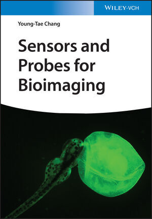
Systematically summarizes the bioimaging chemical probes from chemical points of views.
1 INTRODUCTION OF BIOIMAGING
1 INTRODUCTION OF BIOIMAGING
1.1. Color
1.2 Colorful material
1.3 Light source of bioimaging
1.4 Subcellular imaging
1.5 Cell selective imaging
1.6 Tissue and organ imaging
1.7 Whole body imaging
1.8 Probes in bioimaging
2. CHEMICAL SENSORS AND PROBES FOR BIOIMAGING
2.1 History of dyes in biological stains
2.2 Blood cell staining
2.3 Bacteria staining using Gram method
2.4 Fluorescent sensors and probes
2.5 Representative fluorescent compounds for bioimaging
3. ORGANELLE SELECTIVE PROBES
3.1 Cell plasma membrane
3.2 Endosome and lysosome
3.3 Nucleus and DNA
3.4 Nucleolus and RNA
3.5 ER and Golgi body
3.6 Mitochondria
3.7 Lipid droplet
3.8 Peroxisome
3.9 Cytosol
3.10 Extracellular vesicle
3.11 Nonmembrane-bound condensate
3.12 Organelle probes in live cells and fixed cells
3.13 Modeling for organelle selective probes
4. LIVE CELL SELECTIVE PROBES
4.1. Protein Oriented Live Cell Distinction (POLD)
4.2. Carbohydrate Oriented Live Cell Distinction (COLD)
4.3. Lipid Oriented Live Cell Distinction (LOLD)
4.4. Gating Oriented Live Cell Distinction (GOLD)
4.5. Metabolism Oriented Live Cell Distinction (MOLD)
5. EX-VIVO TISSUE SELECTIVE PROBES
5.1 Immunohistochemistry
5.2 Tissue imaging with nucleic acid probes
5.3 Tissue imaging with small molecule probes
5.3.1 Pancreatic islet imaging
5.3.2 Neuronal tissue imaging
5.4 Organoid as model of tissue and organ
6. IN-VIVO WHOLE-BODY IMAGING PROBES
6.1 ElaNIR for elastin imaging in mouse
6.2 Probes for exposed neuron in zebrafish embryo
6.3 NeuO for whole-body neuron imaging in zebrafish
6.4 LipidGreen for fatty tissue imaging in zebrafish
6.5 Blood vessel imaging in zebrafish
6.6 Probes for bone imaging
6.7 Probes for pancreatic islet imaging
6.8 Probes for eye imaging
6.9 Macrophage imaging in ischemia and inflammation
7. IMAGING FOR BIOLOGICAL ENVIRONMENT AND FUNCTION
7.1 pH
7.2 Metal ions
7.3 Metabolites
7.4 Viscosity
7.5 Temperature
7.6 ROS and RNS
8. IMAGING FOR DISEASE
8.1 Cancer imaging
8.2 Neurodegenerative disease imaging
8.3 Inflammation imaging
8.4 Diabetes imaging
8.5 Liver disease imaging
8.6 Aging
8.7 Theranostics
9. NON-OPTICAL IMAGING PROBES
9.1 Ultrasound imaging probes
9.2 X-ray contrast agents
9.3 MRI contrast agents
9.4 SPECT probes
9.5 PET probes
9.6 Multimodality
10. FLUORESCENCE IMAGING TECHNIQUES AND ANALYSIS METHODS
10.1 Multi-color imaging
10.2 Ratiometric measurement
10.3 Fluorescence lifetime imaging microscopy
10.4 Confocal fluorescence microscopy
10.5 Two photon excitation fluorescence imaging and harmonic generation
10.6 Selective plane illumination microscopy
10.7 Total internal reflection fluorescence microscopy
10.8 Super resolution imaging
10.9 Single molecule imaging
10.10 Photoactivation of caged molecule
10.11 Fluorescence recovery after photobleaching
10.12 Flow cytometry technique
11. PERSPECTIVES FOR FUTURE PROBE DEVELOPMENT
11.1 Design of selective probes
11.2 Discovery of selective probes by screening
11.3 Future probe development
Chang, Young-Tae
Kang, Nam-Young
| ISBN | 9783527349609 |
|---|---|
| Artikelnummer | 9783527349609 |
| Medientyp | Buch |
| Auflage | 1. Auflage |
| Copyrightjahr | 2023 |
| Verlag | Wiley-VCH |
| Umfang | 368 Seiten |
| Abbildungen | 3 SW-Abb., 6 Farbabb., 3 Tabellen |
| Sprache | Englisch |

