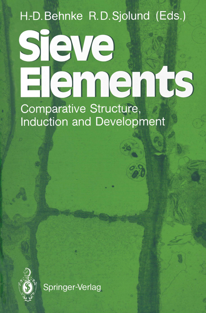
Sieve Elements
Comparative Structure, Induction and Development
Sieve Elements
Comparative Structure, Induction and Development
As part of his Comparative Investigations of the Organization of the Trunk of the Native Forest Trees (Theodor Hartig 1837, Vergleichende Untersuchungen tiber die Organisation des Stammes der einheimischen Waldbaume. lahresberichte tiber die Fortschritte der Forstwissenschaften und forstlichen Naturkunde 1: 125-168) Hartig gives an anatomical description of the "composition and nature" of the then "completely uninvestigated elementary organs" of what he called the "sap skin" (Safthaut) of trees, a tissue for which Nageli later (1858) coined the term phloem. Within the "Safthaut" Hartig describes three cell types in detail, (1) "Siebfasern", (2) "Siebrohren", and (3) "keulenfOrmige Saftrohren" (club-shaped sap-tubes). While the description of the latter refers to laticifers in Euphorbia and resin ducts in Acer and Robinia. "Siebfasern" and "Siebrohren" comprise the sieve elements. A literal translation of the more significant parts of the description of these cell types demonstrates that his "Siebrohren" entirely correspond to what has later been defined as "sieve tubes" but that his "Siebfasern" are less well defined and in addition to what we call "sieve cells" also include small sieve tubes as well as spindle-shaped cells of cambium, phloem parenchyma and sclerenchyma. Both in his "Siebfasern" and "Siebrohren" Hartig describes sieve areas (his expression is "lense-shaped cavities") and sieve pores (Siebporen).
1.2 Medium-Distance Transport
1.3 Long-Distance Transport
1.4 Conducting Cells of Red Algae
1.5 Conducting Cells in Brown Algae
2 Mosses
2.1 Introduction
2.2 General Organization of Conducting Tissues in Mosses
2.3 Structure of Sieve Elements
2.4 Associated Parenchyma
3 Seedless Vascular Plants
3.1 Introduction
3.2 The Sieve-Element Protoplast
3.3 The Wall
3.4 The Sieve Areas
3.5 Parenchymatous Cells Associated with the Sieve Elements
3.6 Longevity of the Sieve Elements
3.7 Comments on Terminology
4 Conifers
4.1 Introduction
4.2 General Description
4.3 Development of the Sieve Cell
4.4 Strasburger Cells
5 Cycads and Gnetophytes
5.1 Introduction
5.2 Cycads
5.3 Gnetophytes
6 Dicotyledons
6.1 Introduction
6.2 The Sieve-Tube Member Protoplast
6.3 The Wall
6.4 The Sieve Plate
6.5 The Lateral Sieve Areas
6.6 Parenchymatous Cells Associated with Sieve-Tube Members
6.7 Longevity of Sieve-Tube Members
7 Monocotyledons
7.1 Introduction
7.2 Ontogeny
7.3 The Protoplast
7.4 Cell Wall
7.5 Thick-Walled Sieve Elements
7.6 Sieve Plates
8 Sieve Elements in Internodal and Nodal Anastomoses of the Monocotyledon Liana Dioscorea
8.1 Introduction
8.2 The Vascular Construction in the Aerial Stem of Dioscorea
8.3 The Specific Composition of Phloem Anastomoses
8.4 Ultrastructure of the Sieve Elements of Anastomoses
8.5 Parenchymatous Cells Associated with the Sieve Elements of Anastomoses
8.6 Some Physiological Implications of Nodal Anastomoses
9 Sieve Elements in Plant Tissue Cultures: Development, Freeze-Fracture, and Isolation
9.1 Introduction
9.2 Phloem Function in Vitro
9.3 Phloem Development in Callus Tissues
9.4 P-Protein, Callus Phloem and Wounding
9.5 Freeze-Fracture Studies Using Callus Sieve Elements
9.6 Sieve-Area Pores
9.7 The Sieve-Element Reticulum (SER)
9.8 Isolation and Partial Purification of Callus Sieve Elements
9.9 Antibody Formation Against Callus Sieve Elements
10 Wound-Sieve Elements
10.1 Introduction
10.2 Tissue Changes During Wound-Phloem Development
10.3 Cytoplasm of Wound-Sieve Elements
10.4 Symplastic Connections of Wound-Sieve Elements
10.5 Companion Cells
10.6 Comparison Between Wound-and Bundle-Sieve Elements
11 Sieve Elements of Graft Unions
11.1 Introduction
11.2 Grafting Procedure
11.3 Histology and Cytology of the Graft Union
11.4 Function of Phloem Connections in Graft Unions
11.5 Questions Concerning the Mechanism of Sieve-Element Formation in Graft Unions
12 Sieve Elements in Haustoria of Parasitic Angiosperms
12.1 Introduction
12.2 Phloem in the Haustorium of Cuscuta
12.3 Development of Haustorial Sieve Elements
12.4 The Contact Hypha of Cuscuta
12.5 Phloem in the Haustoria of Different Parasitic Plants
12.6 Comparative Aspects
13 Phloem Proteins
13.1 Introduction
13.2 P-Protein
13.3 Other Phloem-Specific Proteins
13.4 Biochemistry of Phloem Proteins
14 Phloem Evolution: An Appraisal Based on the Fossil Record
14.1 Introduction
14.2 Phloem of Vascular Cryptogams
14.3 Gymnosperm Phloem
14.4 Conclusions - Phloem Phylogeny.
1 Algae
1.1 Requirement for Medium-Distance and Long-Distance Transport in Algae1.2 Medium-Distance Transport
1.3 Long-Distance Transport
1.4 Conducting Cells of Red Algae
1.5 Conducting Cells in Brown Algae
2 Mosses
2.1 Introduction
2.2 General Organization of Conducting Tissues in Mosses
2.3 Structure of Sieve Elements
2.4 Associated Parenchyma
3 Seedless Vascular Plants
3.1 Introduction
3.2 The Sieve-Element Protoplast
3.3 The Wall
3.4 The Sieve Areas
3.5 Parenchymatous Cells Associated with the Sieve Elements
3.6 Longevity of the Sieve Elements
3.7 Comments on Terminology
4 Conifers
4.1 Introduction
4.2 General Description
4.3 Development of the Sieve Cell
4.4 Strasburger Cells
5 Cycads and Gnetophytes
5.1 Introduction
5.2 Cycads
5.3 Gnetophytes
6 Dicotyledons
6.1 Introduction
6.2 The Sieve-Tube Member Protoplast
6.3 The Wall
6.4 The Sieve Plate
6.5 The Lateral Sieve Areas
6.6 Parenchymatous Cells Associated with Sieve-Tube Members
6.7 Longevity of Sieve-Tube Members
7 Monocotyledons
7.1 Introduction
7.2 Ontogeny
7.3 The Protoplast
7.4 Cell Wall
7.5 Thick-Walled Sieve Elements
7.6 Sieve Plates
8 Sieve Elements in Internodal and Nodal Anastomoses of the Monocotyledon Liana Dioscorea
8.1 Introduction
8.2 The Vascular Construction in the Aerial Stem of Dioscorea
8.3 The Specific Composition of Phloem Anastomoses
8.4 Ultrastructure of the Sieve Elements of Anastomoses
8.5 Parenchymatous Cells Associated with the Sieve Elements of Anastomoses
8.6 Some Physiological Implications of Nodal Anastomoses
9 Sieve Elements in Plant Tissue Cultures: Development, Freeze-Fracture, and Isolation
9.1 Introduction
9.2 Phloem Function in Vitro
9.3 Phloem Development in Callus Tissues
9.4 P-Protein, Callus Phloem and Wounding
9.5 Freeze-Fracture Studies Using Callus Sieve Elements
9.6 Sieve-Area Pores
9.7 The Sieve-Element Reticulum (SER)
9.8 Isolation and Partial Purification of Callus Sieve Elements
9.9 Antibody Formation Against Callus Sieve Elements
10 Wound-Sieve Elements
10.1 Introduction
10.2 Tissue Changes During Wound-Phloem Development
10.3 Cytoplasm of Wound-Sieve Elements
10.4 Symplastic Connections of Wound-Sieve Elements
10.5 Companion Cells
10.6 Comparison Between Wound-and Bundle-Sieve Elements
11 Sieve Elements of Graft Unions
11.1 Introduction
11.2 Grafting Procedure
11.3 Histology and Cytology of the Graft Union
11.4 Function of Phloem Connections in Graft Unions
11.5 Questions Concerning the Mechanism of Sieve-Element Formation in Graft Unions
12 Sieve Elements in Haustoria of Parasitic Angiosperms
12.1 Introduction
12.2 Phloem in the Haustorium of Cuscuta
12.3 Development of Haustorial Sieve Elements
12.4 The Contact Hypha of Cuscuta
12.5 Phloem in the Haustoria of Different Parasitic Plants
12.6 Comparative Aspects
13 Phloem Proteins
13.1 Introduction
13.2 P-Protein
13.3 Other Phloem-Specific Proteins
13.4 Biochemistry of Phloem Proteins
14 Phloem Evolution: An Appraisal Based on the Fossil Record
14.1 Introduction
14.2 Phloem of Vascular Cryptogams
14.3 Gymnosperm Phloem
14.4 Conclusions - Phloem Phylogeny.
| ISBN | 978-3-642-74447-1 |
|---|---|
| Artikelnummer | 9783642744471 |
| Medientyp | Buch |
| Auflage | Softcover reprint of the original 1st ed. 1990 |
| Copyrightjahr | 2012 |
| Verlag | Springer, Berlin |
| Umfang | XIII, 305 Seiten |
| Abbildungen | XIII, 305 p. |
| Sprache | Englisch |

