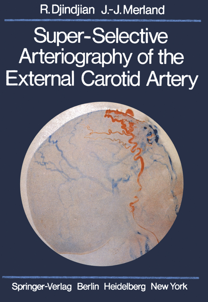
Super-Selective Arteriography of the External Carotid Artery

Super-Selective Arteriography of the External Carotid Artery

235,39 €*

in Vorbereitung

It was a great honour and a mark of our friendship that RENE DJINDJIAN asked me to present this new work from the Lariboisiere neuroradiological school. Professor DJINDJIAN died on 11th October. Introducing this book now, I feel that I am dealing with a fatherless child, where feelings of admiration and pleasure are mixed with the sadness of the loss and the bitterness of an unfinished task. This work on the superselective angiography of the branches of the external carotid artery is a direct continuation of the previous studies of Professor DJINDJIAN, whose name will continue to be closely associated with the development and progress of arteriography in recent years, particularly with arteriography of the spinal cord. It was realised together with his pupil, JEAN-JACQUES MERLAND, whose remarkable thesis for the Doctorat en Medecine a few years ago also dealt with this subject, and with the collaboration of JACQUES THERON, also for many years a pupil of RENE DJINDJIAN, and is translated here by his friend I.F. MOSELEY.

1 Normal Super-Selective Arteriography of the External Carotid Artery
I. Technique of Opacification
A. Techniques of Exploration
1. Femoral Approach
2. Carotid Approach
3. Temporal Approach
B. Conduct of the Examination
C. Injection for Radiography
II. Lingual Artery
A. Technique of Investigation and Projections
B. Morphology of the Lingual Artery
1. Origin of the Lingual Artery
2. Trunk of the Lingual Artery
a) Calibre
b) Carotid and Pharyngeal Segments of the Lingual Artery
c) Lateral Suprahyoid Segment
3. Termination of the Lingual Artery: Deep Lingual Artery
C. Branches of the Lingual Artery
1. Branches of the Pharyngeal and Suprahyoid Segments
2. The Sublingual Artery
3. Branches of the Termination of the Lingual Artery
D. Venous Return
E. Anastomoses
III. Facial Artery
A. Trunk of the Facial Artery
1. Lateral View
a) Initial Horizontal Submental Segment
b) Superficial Segment
2. Antero-Posterior Projection
3. Basal Projection
4. Variations
5. Venous Return
B. Branches of the First Horizontal Segment of the Facial Artery
1. Ascending Palatine Artery
2. Arteries of the Submandibular Gland
a) Main Artery of the Submandibular Gland
b) Accessory Arteries of the Submandibular Gland
3. Submental Artery
C. Branches of the Second, Superficial Segment of the Facial Artery
1. Arteries to the Masseter
2. Genial Arteries
3. Mental Arteries
4. Labial Arteries
a) Inferior Labial (Inferior Coronary) Artery
b) Superior Labial Artery
c) Peri-Oral and Labial Arterial Circle
D. Blood Supply of the Nasal Region and Termination of the Facial Artery
IV. Ascending Pharyngeal Artery
A. Trunk of the Ascending Pharyngeal Artery
1. Lateral Projection
2. Other Projections
B. Branches of the Ascending Pharyngeal Artery
1. Pharyngeal Branches
2. Branches to the Paravertebral Muscles
3. Meningeal and Other Intracranial Branches
4. Tympanic Branches
5. Nervous Branches
C. Anastomoses
V. Occipital Artery
A. Trunk of the Occipital Artery
1. Lateral Projection
2. Frontal (Towne's) Projection
B. Branches of the Occipital Artery
1. Branches of the First Ascending Segment
a) Superior Sterno-Mastoid Artery
b) Stylomastoid Artery
2. Branches of the Second, Horizontal Segment
3. Branches of the Third Segment of the Occipital Artery
C. Venous Return
D. Anastomoses
VI. Posterior Auricular Artery
VII Superficial Temporal Artery
A. Trunk of the Superficial Temporal Artery
1. Lateral Projection
2. Antero-Posterior Projection
B. Branches of the Superficial Temporal Artery
1. Parotid Gland Branches
2. Transverse Facial Artery
3. Zygomaticomalar Artery
4. Anterior Auricular Artery
5. Posterior Deep Temporal Artery
6. Terminal Branches of the Superficial Temporal Artery
C. Venous Return
VIII. Internal Maxillary Artery
A. Trunk of the Internal Maxillary Artery
1. Origin
2. First Segment
3. Second (Zygomatic or Pterygoid) Segment
4. Third, Pterygopalatine Segment
5. Branches of the Internal Maxillary Artery
B. Ascending Cranial and Extracranial Branches of the Internal Maxillary Artery
1. Anterior Tympanic Artery
2. Accessory Meningeal Artery
C. Ascending Extracranial Muscular Branches of the Maxillary Artery
1. Middle Deep Temporal Artery
2. Anterior Deep Temporal Artery
D. Recurrent Branches of the Termination of the Internal Maxillary Artery
1. Vidian Artery or Artery of the Pterygoid Canal
2. Pharyngeal Branch of the Internal Maxillary or Pterygopalatine Artery
3. Artery of the Foramen Rotundum
E. Descending Branches of the Internal Maxillary Artery
1. Masseteric and Pterygoid Branches
2. Inferior Dental Artery
3. Buccal Artery
F. Anterior Branches of the Internal Maxillary Artery
1. Posterior Superior Dental Artery
2. Infraorbital Artery
3. Descending or Superior Palatine Artery
G. Sphenopalatine Artery
1. Origin
2. External Branch: Posterior Lateral Nasal or Lateral Sphenopalatine Artery
3. Posterior Septal, Nasal Palatine, or Medial Sphenopalatine Artery
H. Middle Meningeal Artery and Its Branches
1. Arterial Trunk
2. Branches of the Middle Meningeal Artery
a) Extracranial Branches
b) Basal Branches
c) Anterior Branches
d) Posterior Branches
e) Terminal Branches
3. Territory of the Middle Meningeal Artery
4. Anastomoses With Other Arteries
5. Capillary Phase of Selective Middle Meningeal Arteriography. "Durography"
6. Venous Drainage of the Meninges
7. Conclusion
2 Cervico-Cephalic Vascular Territories
I. Superficial Cutaneous Territories of the Face
A. Supply of the Face
1. Submental Territory
2. Mental Territory
3. Blood Supply of the Lips
4. Nasal Territory
5. Blood Supply of the Cheek
a) Branches of the Facial Artery
b) Branches of the Transverse Facial Artery
c) Branches of the Infraorbital Artery
d) Branches of the Buccal Artery
6. Masseteric Region
a) Masseteric Branches of the Facial Artery
b) Masseteric Branches of the Transverse Facial Artery
c) Masseteric Branches of the Internal Maxillary Artery
7. Zygomaticomalar Region
a) Feeding Arteries
b) Anastomotic Pathways
B. Anastomotic Arcades in the Facial Region
1. Vertical Anastomotic Pathways
a) Postero-Lateral Anastomoses
b) Antero-Lateral Anastomoses
c) Anterior Anastomoses
2. Horizontal Anastomotic Pathways
a) Mandibular and Symphyseal Region
b) Facio-Labial Region
c) Nasal Region
d) Frontal and Glabellar Regions
C. Vascularisation of the Scalp
D. Vascularisation of the Pinna
II. Mucosal Territories
A. Supply of the Buccal Cavity
B. Soft Palate and Pillars of the Fauces
C. Vascularisation of the Oropharynx
D. Blood Supply of the Nasopharynx or Cavum Oris
E. Blood Supply of the Nasal Fossae
1. Nasal Septum
2. Conchae, Meatuses, and Ethmoid Regions
F. Blood Supply of the Maxillary Antra
G. Supply of the Alveolus and Teeth
III. Meningeal and Osseous Territories
A. Supply of the Cranial Vault and Associated Meninges
B. Vascularisation of the Falx Cerebri and the Tentorium
C. Supply of the Skull Base
1. Exocranial Supply
2. Endocranial Supply
a) Squamo-Occipital Portion
b) Meninges of the Petrous Portion of the Posterior Fossa
c) Meningeal Supply of the Clivus
d) Supply of the Middle Cranial Fossa
e) Meningeal Supply of the Anterior Cranial Fossa
IV. Orbital Territory
3 Super-Selective External Carotid Angiography in Pathological Conditions Cranio-Facial Angiomas
I. General Principles, Embolisation
II. Cranio-Facial Angiomas of the External Carotid Territory
A. General Considerations
B. Arteriovenous Fistulae
1. Morphological Data
2. Embolisation of Angiomas With Shunts or Fast Flow Angiomas
3. Morphology and Embolisation of the Arteriovenous Angiomas
a) Localised Arteriovenous Fistulae Supplied Only by the External Carotid Artery
b) Regional Arteriovenous Fistulae Supplied Exclusively by the External Carotid Artery
c) Arteriovenous Fistulae, in Which the External Carotid Artery or Its Branches Have Been Tied
d) Arteriovenous Fistulae, Small or Large, Receiving Supply From the Internal Carotid Artery
e) Large Angiomas in the Territory of Both Internal and External Carotid Arteries
C. Capillary Angiomas
1. Morphology
2. Technique of Injection
3. Embolisation and Intra-Arterial Treatment of Capillary Angiomas
D. Venous Angiomas
1. Morphology
2. Intra-Arterial Treatment of Venous Angiomas
E. Mixed Type Angiomas
F. Intra-Arterial Treatment of the Complications of Angiomas
4 Tumours Supplied by the External Carotid Artery. Super-Selective Injection of the External Carotid Artery in the Investigation of Tumours
I. Meningiomas
A. Convexity Meningiomas
1. Feeding Vessels
a) Parasagittal Meningiomas
b) Supratentorial Convexity Meningiomas
c) Infratentorial Convexity Meningiomas
2. Tumour Vascularity
3. Osseous and Superficial Extension
4. Draining Veins
5. Invasion of the Veins
6. Embolisation
B. Tentorium
C. Base of the Skull
II. Acoustic Neuromas
III. Glomus Tumours
1. Feeding Vessels
2. Tumour Vascularity
3. Venous Drainage
4. Embolisation
IV. Other Intracranial Tumours
V. Sturge Weber Syndrome and Migraine
VI. Tumours and Other Diseases of the Cranial Vault
VII. Tumours in Ear, Nose and Throat and Facio-M axillary Surgery
5 Meningeal Arteriovenous Fistulae
I. Pure Meningeal Arteriovenous Fistulae
A. General Data
1. Historical Aspects
2. Classification
3. Protocol of Angiographic Investigation
B. Morphology of Pure Meningeal Arteriovenous Fistulae
1. Pure Meningeal Arteriovenous Fistulae, Draining Directly into a Venous Sinus or Meningeal Vein
a) Pure Meningeal Arteriovenous Fistulae in the Wall of the Lateral Sinus
b) Pure Arteriovenous Fistulae of the Walls of the Cavernous Sinus or of the Neighbouring Meninges, Draining into the Cavernous Sinus
c) Pure Arteriovenous Fistulae of the Superior Sagittal Sinus and the Convexity
d) Pure Dural Arteriovenous Fistulae With Mixed Venous Drainage into the Sinus and Local Veins
2. Pure Meningeal Arteriovenous Fistulae Draining to Cortical Veins
a) Clinical Features
b) Morphological and Arteriographic Features
c) Unique Features of the Venous Drainage
d) Therapeutic Considerations
3. Meningeal Arteriovenous Fistulae With Large Dural Venous Lakes
II. Mixed Meningeal Arteriovenous Fistulae
A. Cutaneous, Bony and Meningeal Arteriovenous Fistulae
B. Meningeal/Cortical Arteriovenous Fistulae
Conclusions.
ISBN 978-3-642-66598-1
Artikelnummer 9783642665981
Medientyp Buch
Auflage Softcover reprint of the original 1st ed. 1978
Copyrightjahr 2011
Verlag Springer, Berlin
Umfang XVIII, 554 Seiten
Abbildungen XVIII, 554 p.
Sprache Englisch