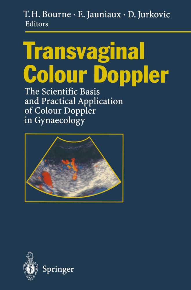
Transvaginal Colour Doppler
The Scientific Basis and Practical Application of Colour Doppler in Gynaecology
Transvaginal Colour Doppler
The Scientific Basis and Practical Application of Colour Doppler in Gynaecology
Since the pioneering work of Donald and his first Lancet paper in 1958, the use of ultrasound in obstetrics and gynaecology has evolved rap idly. The introduction of grey scale techniques enhanced our ability to identify different tissues on the basis of their texture. However, it was the introduction of the linear array real-time scanner in the mid seven ties that changed ultrasound from being an "eccentric art form" to a readily available and usable technique. This led to the first reports of the diagnosis of neural tube defects using ultrasound by Campbell, as well as the establishment of fetal biometry. In the midst of this activity the parallel development of the transvaginal probe by Kratochwill went almost unnoticed by most gynaecologists. Yet the application of this technique has since had a major impact on many areas of gyna ecological practice, and on infertility in particular. Since the demon stration of transvaginal follicle aspiration, the vaginal route has become standard for most invasive ultrasound guided gynaecological procedures. The relatively new technical advance of transvaginal colour Doppler may potentially have just as great an impact. The introduction and use of transvaginal colour flow imaging has facili tated the study of vascular changes within the pelvis.
Angiogenesis
Vascular Anatomy of the Pelvis
The Human Heart - Development of Form and Function
Vascular Features of Gynaecological Neoplasms
Vascular Physiology and Pathophysiology of Early Pregnancy
New Diagnostic and Therapeutic Approaches to Gestational Trophoblastic Tumors
II. Uterus
Vascular Changes During the Normal and Artificial Cycle
Uterine and Endometrial Pathology
Investigation of the Utero-Placental Circulation
Transvaginal Colour Doppler in Trophoblastic Disease
III. Ovaries and Fallopian Tubes
Vascular Changes During the Ovarian Cycle
Ovulation and the Periovulatory Follicle
The Study of Ovarian Tumours
Transvaginal Color Doppler Ultrasound in Pelvic Inflammatory Disease
Transvaginal Colour Doppler Studies of Ectopic Pregnancy
Hystero-Contrast-Salpingography - Colour Doppler and the Study of Fallopian Tube Patency
IV. Embryo or Fetus
Assessment of the Early Foetal Circulation
Transvaginal Examination of the Foetal Heart
V. Urinary Tract
Doppler Studies of the Lower Urinary Tract.
I. General Principles
Principles of Colour DopplerAngiogenesis
Vascular Anatomy of the Pelvis
The Human Heart - Development of Form and Function
Vascular Features of Gynaecological Neoplasms
Vascular Physiology and Pathophysiology of Early Pregnancy
New Diagnostic and Therapeutic Approaches to Gestational Trophoblastic Tumors
II. Uterus
Vascular Changes During the Normal and Artificial Cycle
Uterine and Endometrial Pathology
Investigation of the Utero-Placental Circulation
Transvaginal Colour Doppler in Trophoblastic Disease
III. Ovaries and Fallopian Tubes
Vascular Changes During the Ovarian Cycle
Ovulation and the Periovulatory Follicle
The Study of Ovarian Tumours
Transvaginal Color Doppler Ultrasound in Pelvic Inflammatory Disease
Transvaginal Colour Doppler Studies of Ectopic Pregnancy
Hystero-Contrast-Salpingography - Colour Doppler and the Study of Fallopian Tube Patency
IV. Embryo or Fetus
Assessment of the Early Foetal Circulation
Transvaginal Examination of the Foetal Heart
V. Urinary Tract
Doppler Studies of the Lower Urinary Tract.
Bourne, Tom H.
Jauniaux, Eric
Jurkovic, Davor
Athanasiou, S.
Bauer, B.
Bicknell, R.
| ISBN | 978-3-642-79266-3 |
|---|---|
| Artikelnummer | 9783642792663 |
| Medientyp | Buch |
| Auflage | Softcover reprint of the original 1st ed. 1995 |
| Copyrightjahr | 2012 |
| Verlag | Springer, Berlin |
| Umfang | XII, 206 Seiten |
| Abbildungen | XII, 206 p. 39 illus. in color. |
| Sprache | Englisch |

