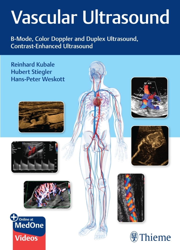
Vascular Ultrasound
An interdisciplinary guide to color duplex sonography organized by anatomic region
The indications for vascular color duplex sonography (CDS) have expanded in recent years due to the availability of power Doppler, B-flow, ultrasound contrast agents, 3D reconstruction techniques and fusion with other imaging modalities. CDS enables close-interval follow-ups after interventional procedures with improved prognoses. Edited by Reinhard Kubale, Hubert Stiegler, and Hans-Peter Weskott, Vascular Color Duplex Ultrasound starts with the basic principles of diagnostic ultrasound physics and technology, followed by invaluable tips on equipment settings, possible artifacts, and limitations; hemodynamic essentials; and the use of ultrasound contrast agents. Subsequent chapters organized by anatomic region provide updated coverage on all peripheral and abdominal arterial and venous vascular regions; microcirculation and tumor perfusion; kidney and liver disease; the use of contrast-enhanced ultrasound (CEUS) in biliary, intestinal, splenic, and pediatric diseases; and novel/future techniques.
Key Features
- Contributions from interdisciplinary experts in angiology, neurology, radiology, vascular surgery, gastroenterology, nephrology, phlebology, rheumatology, laser medicine, and physics
- In-depth guidance on examination techniques, findings, and potential pitfalls and how to avoid them
- A wealth of comparative CT, MRI, and angiography CDS images and 37 videos enhance understanding of impacted anatomy, and the ability to master techniques and make accurate diagnoses
This book includes complimentary access to a digital copy on https://medone.thieme.com
Part I: Basic Principles
1 Principles of Physics and Technology in Diagnostic Ultrasound
2 Ultrasound Device Settings, Examination Technique, and Artifacts
3 Hemodynamics
4 Ultrasound Contrast Agents Fundamentals and Principles of Use
Part II: Vascular Ultrasound
5 Extracranial Cerebral Arteries
6 Intracerebral Arteries and Brain
7 Limbs
8 Nonatherosclerotic Arterial Diseases: Vasculitis, Fibromuscular Dysplasia, Cystic Adventitial Disease, Compression Syndromes
9 Vascular Malformations
Part III: Abdominal Organs: Vascularization and Perfusion
10 Aorta and Outgoing Branches
11 Visceral Arteries
12 Abdominal Veins
13 Microcirculation and Tumor Perfusion
14 Kidneys and Renal Transplants
15 Liver and Portal Venous System
16 Contrast-Enhanced Ultrasound (CEUS) in Biliary Diseases
17 Contrast-Enhanced Ultrasound (CEUS) in Intestinal Diseases
18 Contrast-Enhanced Ultrasound (CEUS) in Pancreatic Diseases
19 Contrast-Enhanced Ultrasound (CEUS) in Splenic Diseases
20 Contrast-Enhanced Ultrasound (CEUS) in Pediatric Diseases
21 Novel and Upcoming Ultrasound Techniques
Kubale, Reinhard
Stiegler, Hubert
Weskott, Hans-Peter
| ISBN | 9783132405431 |
|---|---|
| Artikelnummer | 9783132405431 |
| Medientyp | Non Books |
| Copyrightjahr | 2023 |
| Verlag | Thieme, Stuttgart |
| Umfang | 574 Seiten |
| Abbildungen | Beilage: Video |
| Sprache | Englisch |

